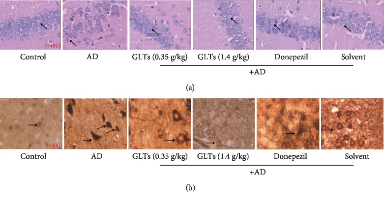
Figure 2.
Effects of GLTs on the hippocampal tissue structure and neuronal tangles of the hippocampus of APP/PS1 transgenic mice. (a) HE staining was applied to present pathological changes in hippocampus tissues of mice in each group (×400). (b) The number of neuronal tangles (NFTs) in the CA1 area of the mouse hippocampus was detected by silver staining in each group (×400), N = 6.

