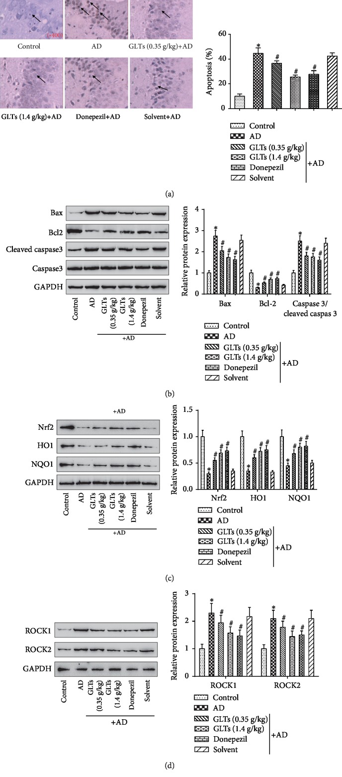
Figure 3.
Effects of GLTs on apoptosis and oxidative damage in hippocampal area of APP/PS1 transgenic mice through ROCK signaling pathway. (a) The number of apoptotic positive cells in the CA1 area of the mouse hippocampus was measured in each group by TUNEL assay (×400). (b) The expression of apoptosis-related protein Bax, Bcl2, and caspase 3/cleaved caspase 3 in each group was detected by western blot. (c) Western blot analysis of the antioxidative proteins Nrf2, HO1, and NQO1 expression levels in the mouse hippocampus. (d) The protein expression of ROCK signaling pathway-associated proteins ROCK1 and ROCK2 in hippocampal tissues was determined. N = 6; ∗P < 0.05 compared to the control group, #P < 0.05 compared to the AD group.

