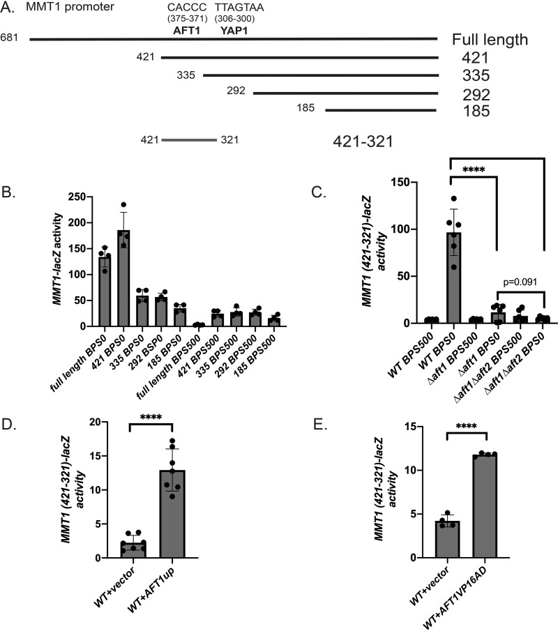
Figure 4.
Aft1 is necessary for low-iron-mediated induction of MMT1. A, diagram of the MMT1 promoter with putative Aft1- and Yap1-binding sites and truncation constructs. B, WT cells transformed with full-length or truncation mutants of the MMT1 promoter fused to lacZ were grown in CM medium with 80 μm BPS (low iron) or high iron (500 μm) FeSO4 overnight. β-Gal activity and protein levels were determined. Error bars represent S.D. n = 4. C, WT, Δaft1, or Δaft1Δaft2 cells expressing MMT1 421–321-lacZ were grown as described in B, and β-gal activity was measured. Error bars represent S.D. n = 6. D, WT cells expressing MMT1 421–321-lacZ were transformed with an empty vector or an AFT1up plasmid. Cells were grown in CM medium overnight, and β-gal activity was measured. Error bars represent S.D. n = 7. E, WT cells expressing MMT1 421–321-lacZ were transformed with an empty vector or an AFT1VP16AD plasmid. Cells were grown in CM medium overnight, and β-gal activity was measured. Error bars represent S.D. n = 4. ****, p ≤ 0.0001.

