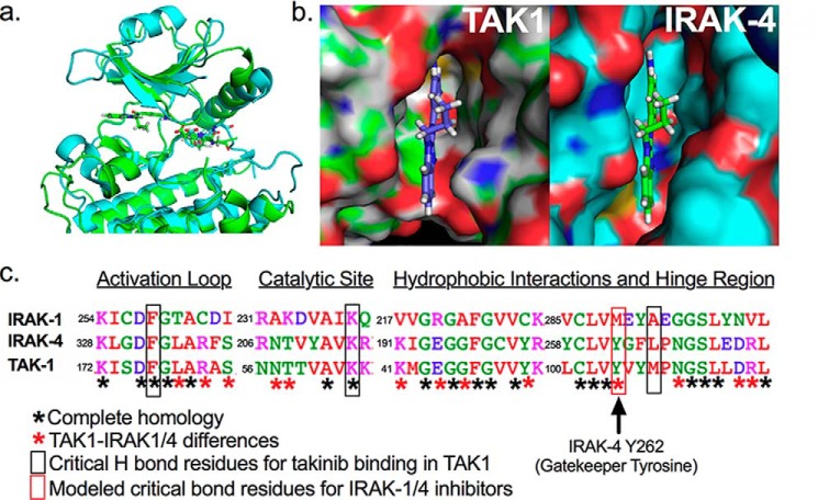
Figure 1.
Structural comparison of TAK1 against IRAK-1 and -4. a, overlay of the ribbon structure of IRAK-4 and TAK1. b, close-up view of the surface representation of takinib in the ATP binding pocket of TAK1 and modeled into IRAK-4. c, amino acid sequence alignment of TAK1, IRAK-4, and IRAK-1 highlighting key amino acid differences.

