Figure 4.
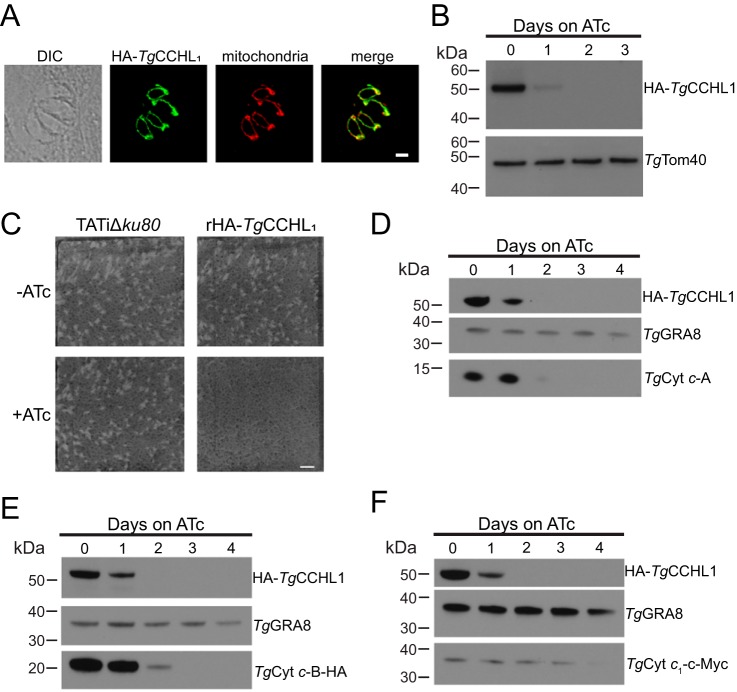
Knockdown of TgCCHL1 results in defects in parasite proliferation and a depletion in the abundance of c-type cytochromes. A, immunofluorescence assay of a four-cell parasite vacuole with HA-TgCCHL1 (green) co-localizing with the mitochondrial marker Tom40 (red). Scale bar is 2 μm. B, Western blots of iHA3-TgCCHL1 lines grown in the presence of ATc for 0–3 days and probed with antibodies against HA and TgTom40 (as a loading control). C, plaque assay of parental TATiΔku80 and rHA-TgCCH1L parasites grown in the absence or presence of ATc over 8 days. Data are from a single experiment and representative of three independent experiments. Scale bar is 10 mm. D–F, Western blots of rHA3-TgCCHL1 parasite strains grown in the presence of ATc for 0–4 days and probed with antibodies against (D) TgCyt c-A, (E) HA-tagged TgCyt c-B, and (F) c-Myc-tagged TgCyt c1. GRA8 is included as a loading control, and data are representative of three independent experiments.

