Figure 4.
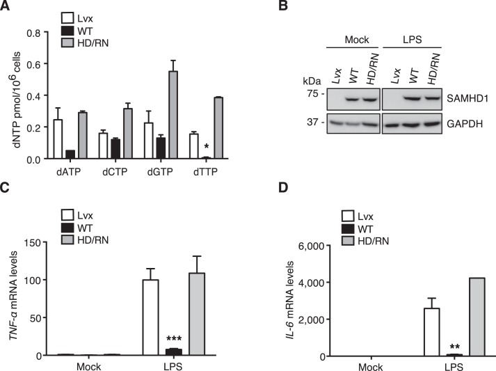
Reconstitution of WT SAMHD1 in differentiated THP-1/KO cells suppresses NF-κB activation. THP-1/KO cells were transduced with a Lvx vector (Lvx), or SAMHD1 WT or HD/RN expressing lentiviruses. The reconstituted THP-1/KO cells were differentiated with PMA (30 ng/ml) for 24 h to induce WT SAMHD1 or HD/RN expression. A, after differentiation, intracellular dNTP levels were measured. Reconstitution of WT SAMHD1, but not HD/RN, in THP-1/KO cells reduces intracellular dNTP levels. Results are shown as mean ± S.D. Statistical significance was determined by unpaired Student's t test; *, p < 0.05 compared with the vector control. B–D, the differentiated cells were treated with LPS (100 ng/ml) or mock treated. After 6-h treatment, cells were collected and cell lysates were analyzed by IB to measure the expression levels of WT SAMHD1 or HD/RN. GAPDH was a loading control (B). Relative mRNA levels of TNF-α (C) and IL-6 (D) in the cell pellets were also measured by RT-PCR. Results are shown as mean ± S.D. Statistical significance was determined using unpaired Student's t test; **, p < 0.01; ***, p < 0.001 compared with vector controls. The results are representative of three independent experiments.

