Figure 5.

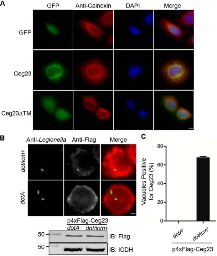
Cellular localization of Ceg23. A, Ceg23 localized to the ER in a manner that requires the two predicted transmembrane domains. HeLa cells were transfected with plasmids that direct the expression of GFP–Ceg23 or GFP–Ceg23ΔTM. The ER was labeled by immunostaining with antibodies specific for the ER-resident protein calnexin. Bar, 5 μm. B, the association of Ceg23 with the LCV. U937 cells infected with the indicated L. pneumophila strains expressing 4× FLAG–Ceg23 were sequentially immunostained with antibodies against L. pneumophila and FLAG. Images were obtained by a fluorescence microscope. The lower panel indicated that 4× FLAG–Ceg23 was similarly expressed in dotA− and dot/icm+ strains. C, quantification of LCVs positively stained for 4× FLAG–Ceg23 shown in B. The results in B and C are from one representative experiment done in triplicate from three independent experiments; at least 100 vacuoles were counted in each sample. Bar, 5 μm. The error bars represent S.E. (n = 3). DAPI, 4′,6′-diamino-2-phenylindole; IB, immunoblot.
