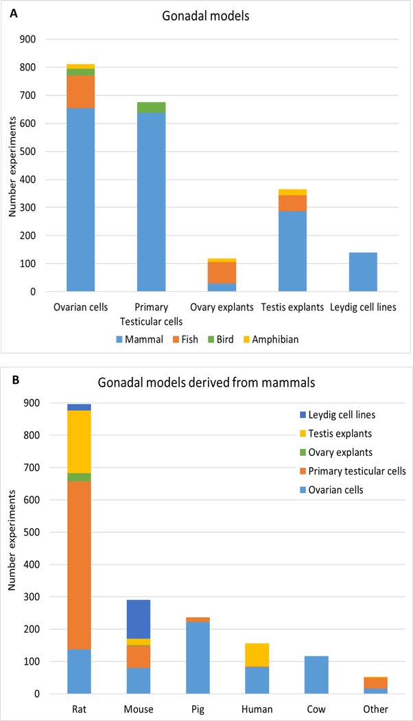
Figure 3. Gonadal models represented in the literature search.
The number of studies indicating the distribution of gonadal models per animal class is represented in A. Ovarian cells include granulosa cells, theca cells, co-cultures and ovarian follicles. Primary testicular cells are represented mostly by purified Leydig cells. Leydig cell lines constitute tumor-derived cells (BLTK-1, R2C, MA-10, MLTC-1) and Leydig cells isolated from normal immature testis (TM-3). Ovary and testis explants constitute whole tissue cultures, fragments and slices. The distribution of gonadal models per mammalian animal sources are indicated in B. “Other” refers to studies using bank vole, hamster, buffalo, monkey, dog, sheep, and rabbit gonadal models.

