Figure 1.

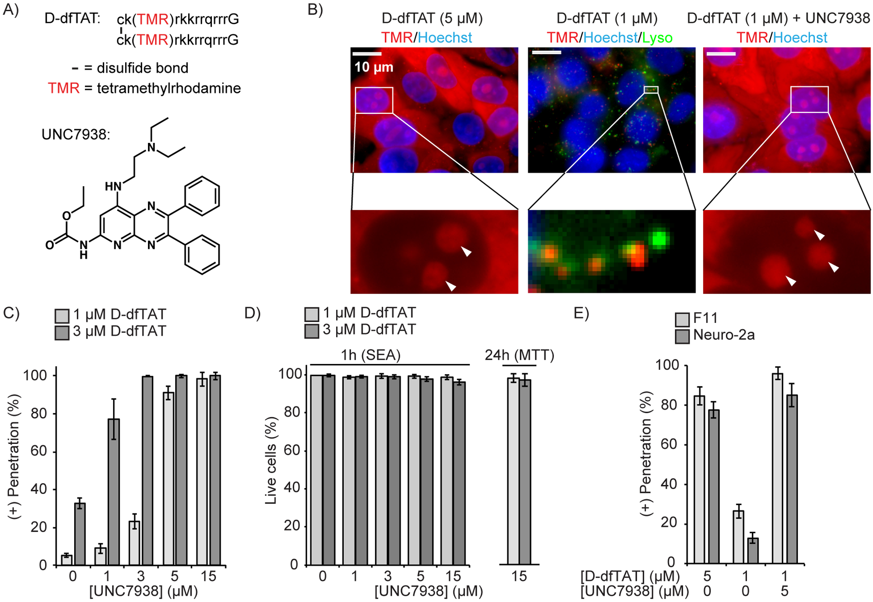
UNC7938 enhances cytosolic penetration of the CPP D-dfTAT. A) Structures of the peptide D-dfTAT and of the small molecule UNC7938. Lower case letters in the peptide sequence refer to D-amino acids. dfTAT has the identical structure but is synthesized with L-amino acids instead. B) Fluorescence microscopy images of HeLa cells treated with D-dfTAT and UNC7938 for 30 min. Images are overlays of fluorescence images pseudo-colored red for TMR and blue for Hoechst 33342. An overlay with Lysotracker Green, pseudo-colored green, is also shown for the condition showing a punctate peptide distribution. Nucleoli are highlighted with white arrows in zoomed-in images. C) HeLa cells were incubated with D-dfTAT and UNC7938 for 30 min, washed, incubated with SYTOX green and Hoechst 33342, and imaged. The cells displaying D-dfTAT positive nucleolar staining while excluding SYTOX green were counted as alive and positive for cell penetration by the peptide. The total number of cells present was established by counting all Hoechst 33342 stained nuclei. The data represented are the means of biological triplicates with corresponding standard deviations (>500 cells counted per experiment). D) Quantification of cell viability 1 and 24 h after incubation with D-dfTAT and UNC7938 (30 min incubation), as measured by a SYTOX Green exclusion assay (SEA) and MTT assay. The data represented correspond to the mean of biological triplicates (>500 cells counted per experiment). E) Effect of UNC7938 on D-dfTAT cell penetration in the F11 and neuro-2a cell lines.
