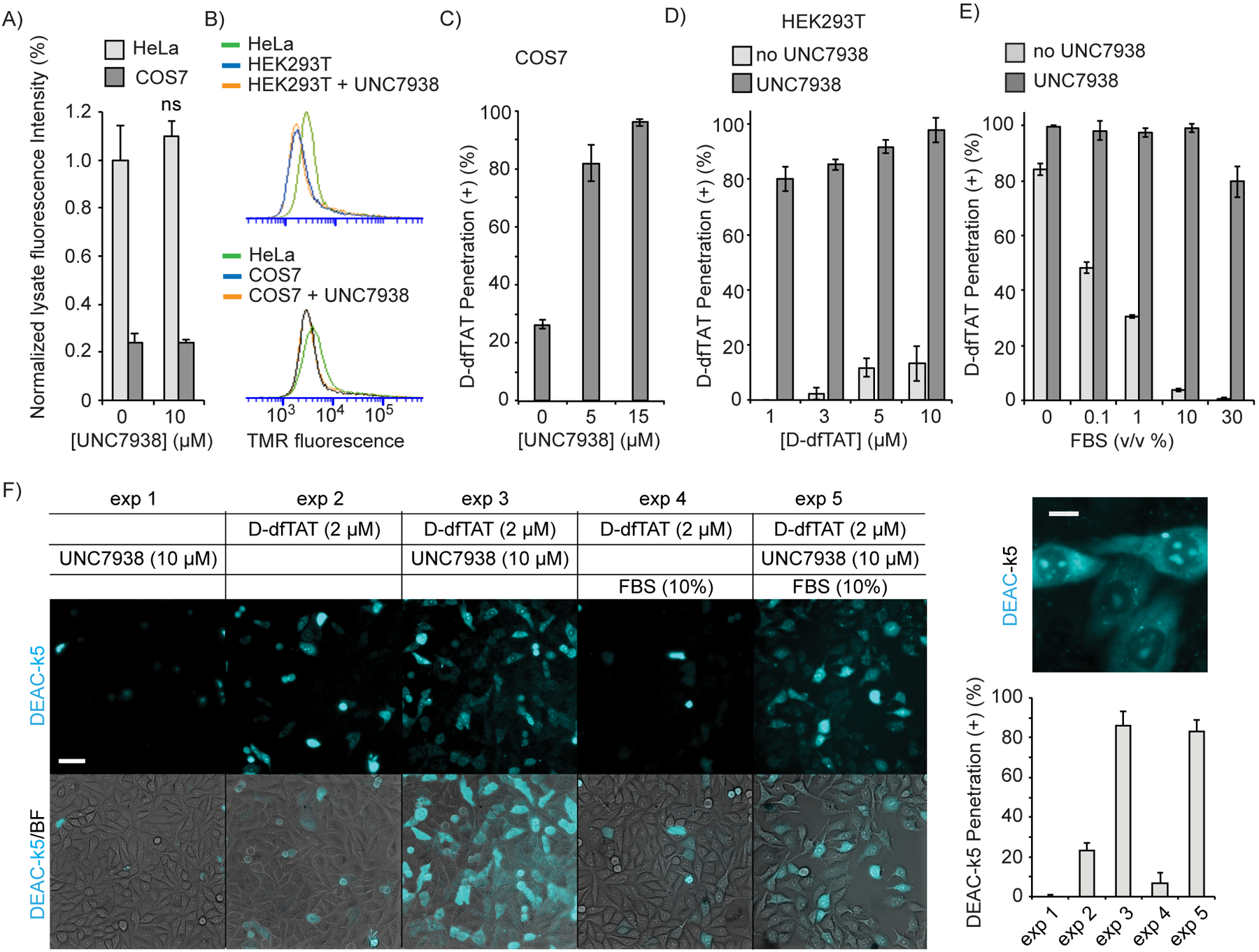
Figure 3.

UNC7938 rescues the cytosolic penetration of D-dfTAT under conditions were CPP alone fail. A) Total uptake of D-dfTAT (5 μM) in HeLa and COS7 cells, with or without UNC7938 after 30 min incubation (ns = not significant by a two-tailed t-test). B) Uptake of TMR-r9 (5 μM) in HeLa, COS7, and HEK293T, as assessed by flow cytometry. Incubation were performed for 30 min, with or without UNC7938 (10 μM). C) Effect of UNC7938 on D-dfTAT penetration in COS7 cells. COS7 cells were treated for 30 min with D-dfTAT (5 μM) and with UNC7938. D) Effect of UNC7938 on D-dfTAT penetration in HEK293T cells. HEK293T cells were incubated for 30 min with D-dfTAT in the absence or presence of UNC7938 (5 μM). E) Effect of FBS on D-dfTAT (5 μM) penetration in HeLa cells, in the presence or absence of UNC7938 (10 μM). F) Delivery of DEAC-k5 in Hela cells with and without FBS. Cells were incubated with DEAC-k5 (10 μM), D-dfTAT (2 μM) and UNC7938 (10 μM) in L15 media with or without 10 % FBS. Representative 20X fluorescence images, pseudo-colored cyan for DEAC and overlaid with bright field images, are shown. A 100X fluorescence image of highlighting the subcellular localization of DEAC-k5 obtained after delivery with D-dfTAT and UNC7938 is provided. The cell penetration of DEAC-k5 is assessed by counting the number of cells displaying nucleolar staining of the peptide. In all panels, the data represented correspond to the mean of biological triplicates (>500 cells counted per experiment). Scale bars 20X: 50 μm, 100X: 10 μm.
