Figure 5.
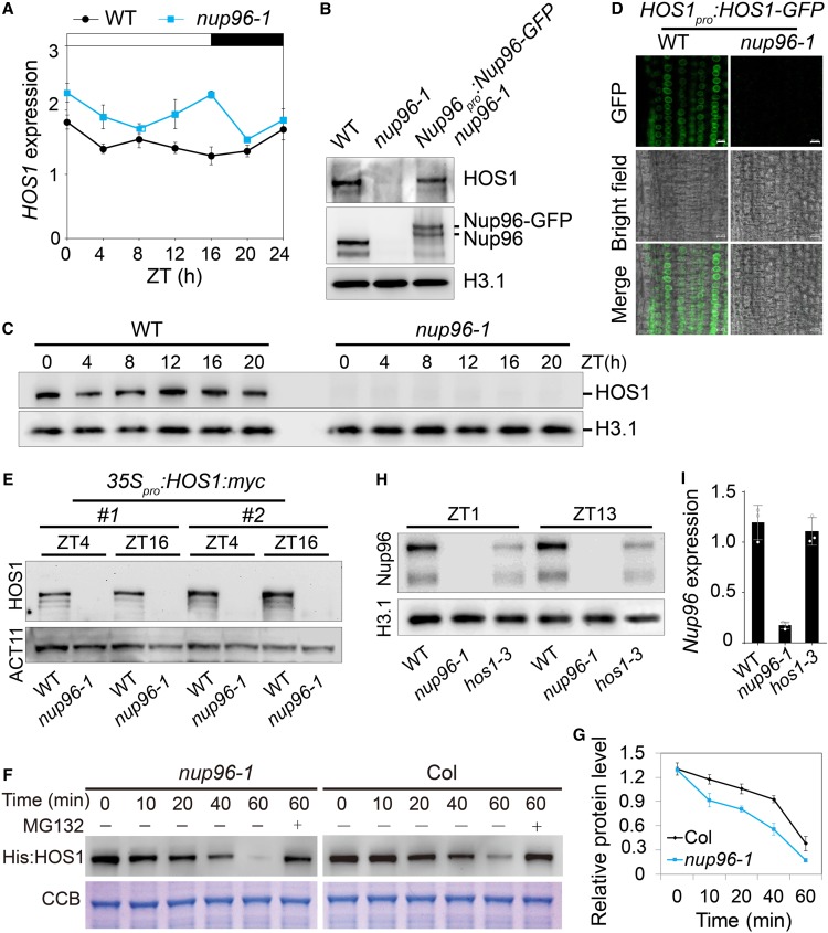
Nup96 Stabilizes HOS1 Proteins in Plants.
(A) Diurnal expression of HOS1 in wild-type (WT) and nup96-1 seedlings grown in long-day conditions for 9 d. Values are means ± sd (n = 3 biological repeats).
(B) Immunoblot shows HOS1 levels in the nuclear extracts from the WT, nup96-1, and Nup96-GFP nup96-1 complementation plants. Histone H3.1 (H3.1) was used as the loading control.
(C) Levels of endogenous HOS1 proteins in the nuclear extracts from WT and nup96-1 seedlings grown in long-day conditions for 9 d, detected by anti-HOS1 antibodies. Histone 3.1 was used as the loading control.
(D) HOS1-GFP signals in wild-type and nup96-1 seedling root cells. Bars = 10 µm.
(E) Levels of HOS1-myc proteins in the nuclear extracts from WT and nup96-1 seedlings grown in long-day conditions for 9 d. ACTIN11 (ACT11) was used as the loading control.
(F) Assay of HOS1 protein stability in vitro. His:HOS1 proteins were purified from Escherichia coli and incubated with the total extracts of nup96-1 or Col-0 seedlings in the presence or absence of MG132. His:HOS1 proteins were probed by His antibody on an immunoblot (upper panel). Coomassie Brilliant Blue (CBB) staining is shown in the lower panel.
(G) Quantitative assay of HOS1 protein levels from three biological repeats with a representative result shown in (F).
(H) Levels of endogenous Nup96 proteins in the nuclear extracts from WT, hos1-3, and nup96-1 seedlings grown in long-day conditions for 9 d.
(I) qPCR analysis of Nup96 expression in WT, hos1-3, and nup96-1 seedlings grown in long-day conditions for 9 d. Values are means ± sd (n = 3 biological repeats).

