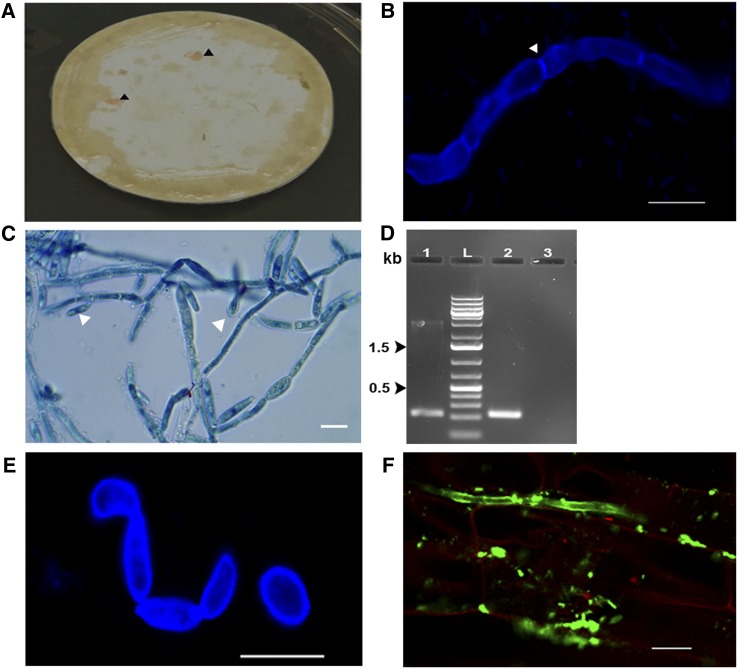
Figure 4.
R. mucilaginosa JGTA-S1 Shows a Transition from Yeast to Filamentous Form.
Rice plants grown under sterile conditions were supplemented with JGTA-S1. Extracts from surface-sterilized plants were passed through a 0.45-µm filter at 3 wpi. The filters were incubated on PDA plates for 3 d at 30°C, followed by 2 weeks or more at 5°C on PDA plates containing antibiotics.
(A) Pink material on filter paper (arrowhead).
(B) Calcofluor-stained JGTA-S1 showing septate hypha (arrowhead).
(C) Lactophenol cotton blue–stained JGTA-S1 filaments with budding yeast-like structures (arrowhead).
(D) Partial D1/D2 sequence amplified from genomic DNA isolated from filamentous JGTA-S1 and compared with the yeast form. Lane 1, amplicon from JGTA-S1 in filamentous form; lane L, DNA markers; lane 2, amplicon from JGTA-S1 in yeast form; lane 3, no DNA control.
(E) Calcofluor staining of JGTA-S1 filament induced by FK506.
(F) Rice roots infected with JGTA-S1 at 9 DAI stained with Alexa Fluor 488–labeled WGA and counterstained with propidium iodide. The roots were imaged under a confocal microscope. Bar = 10 µm

