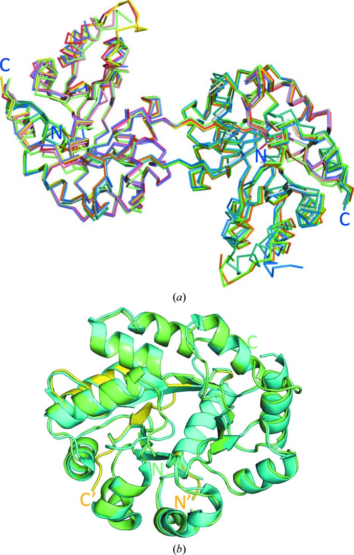
Figure 2.
Comparison of SpTrpA dimers and the TIM-barrel fold. (a) Cα superposition of the five dimers: A/B, red/blue; C/D, yellow/green; E/F, salmon/gray; G/H, violet/orange; I/J, light green/teal. The superposition was calculated using the align procedure in PyMOL. (b) Superposition of the TIM barrel of SpTrpA formed from chains C (residues 1–59, yellow) and D (60–258, green) with the model of the SpTrpA subunit (cyan) from the structure of SpTrpAB (PDB entry 5kin; r.m.s.d. of 0.45 Å on Cα superposition).

