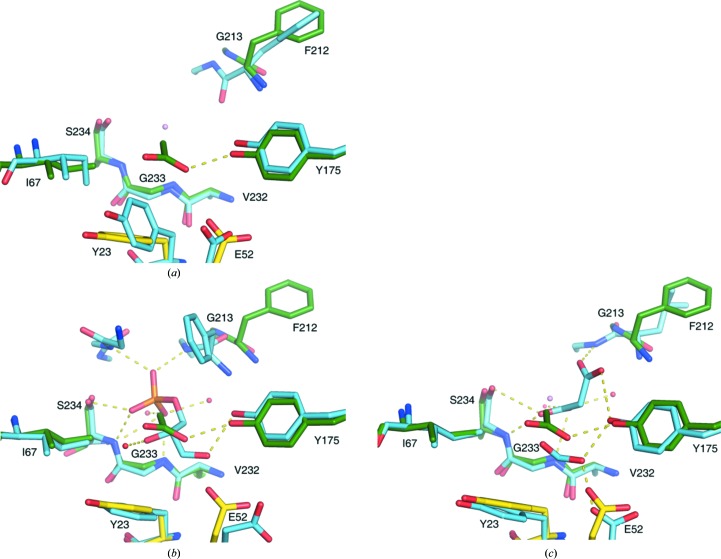
Figure 3.
Details of the SpTrpA active site. The active site of SpTrpA, chain C (yellow) and chain D (green), including the acetate ion (ACY; green) superposed with (a) chain A of the SpTrpAB structure (PDB entry 5kin; blue), (b) chain A of the StTrpA structure (PDB entry 1wbj; blue) including the sn-glycerol-3-phosphate ligand (G3P; blue), with water molecules from the StTrpA structure shown as red spheres, and (c) chain A of the MtTrpAB structure (PDB entry 5tcf; blue) including the formic acid and malonate ligands (FMT and MLI; blue), with water molecules from the MtTrpAB structure shown as red spheres. Selected hydrogen bonds are marked as yellow dashes. A water molecule from the present SpTrpA structure is shown as a pink sphere.

