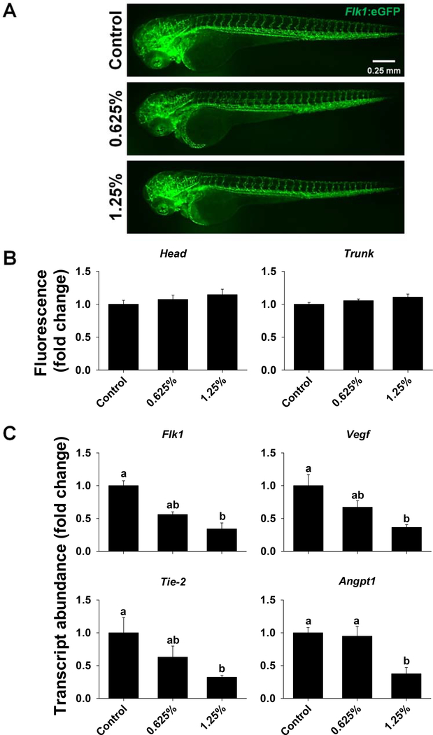
Figure 7.
Vascular development in zebrafish exposed to 0%, 0.625% or 1.25% 1,2-propanediol from 6 hpf until 72 hpf. Tg(Flk1:eGFP) zebrafish were used to visualize the expression of Flk1; fluorescence images of zebrafish were captured at 72 hpf (A), and fluorescent signal in the head and trunk regions was quantified using Image J (B). Transcript abundance of Flk1, Vegf, Tie-2, and Angpt1 was assessed in wild type zebrafish at 72 hpf (C). Letters indicate significant differences that were assessed using one-way ANOVA with a post hoc Tukey test; P ≤ 0.05 was considered significant.

