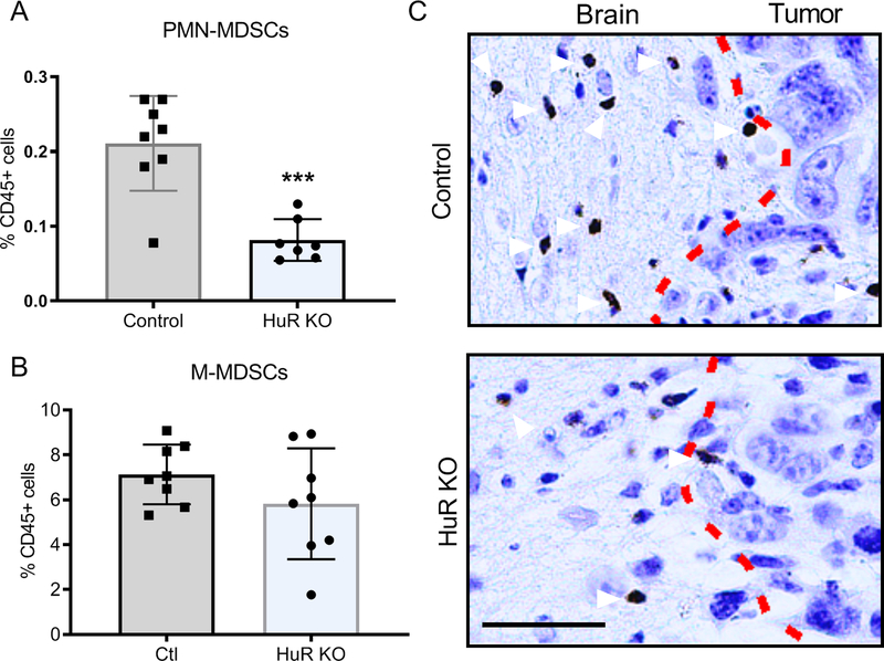
Figure 4. Polymorphonuclear myeloid-derived suppressor cells (PMN MDSCs) are attenuated in HuR KO tumors.
(A) Brain PMN MDSCs (CD45hi, CD11b+,Gr1hi,Ly6Clow, Ly6G+CD49d−) were quantified by flow cytometry. (B) Brain monocytic MDSCs (CD45hiCD11b+Gr1midLy6ChiLy6G−CD49d+) were quantified by flow cytometry. (C) Immunostaining for neutrophils (arrowheads) in brain sections using an MPO antibody. Dotted line represents the border between normal brain and tumor. Each data point is representative of an individual mouse. Means and SDs are shown. ***P = 0.0003. Scale bar, 50 μm.

