Fig. 1.

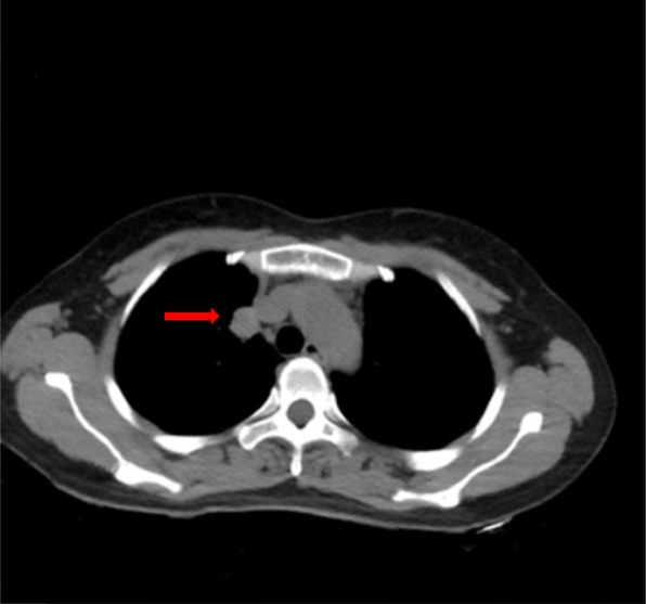
Chest computed tomography showed a 2.1 × 1.7 cm well-defined round mass exhibiting mild, heterogeneous internal enhancement at the periphery of the right superior lobe

Chest computed tomography showed a 2.1 × 1.7 cm well-defined round mass exhibiting mild, heterogeneous internal enhancement at the periphery of the right superior lobe