Fig. 3.
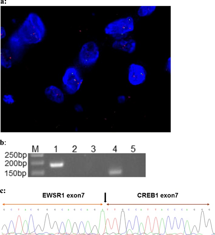
a: Dual color interphase fluorescence in situ hybridization utilizing the EWSR1 break-apart probe. Split red and green signals within a single tumor cell demonstrated the presence of EWSR1 rearrangement. b: Gel electrophoresis of the RT-PCR products using EWSR1 and CREB1 primers; confirming presence of ESWR1-CREB1 fusion in the patient’s sample (Lane 4). M:50 bp markers; Lane 1: Internal control, PGK; Lane 2: Negative control with EWSR1 exon 7 + CREB1 exon 7 fusion primer; Lane 3: Negative control with EWSR1 exon 7 + CREB1 exon 8 fusion primer; Lane 4: Patient’s sample with EWSR1 exon 7 + CREB1 exon 7 fusion primer; Lane 5: Patient’s sample with EWSR1 exon 7 + CREB1 exon 8 fusion primer. c: Sanger sequencing result of the patient’s RT-PCR product demonstrated in Lane 4 of b. The sequence was the same as the EWSR1 - CREB1 fusion gene as reported in the literature

