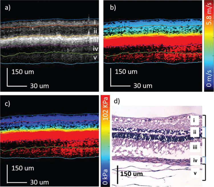
Figure 5.
In-vivo rabbit elastography results. a) OCT of rabbit central retina. b) Shear wave velocity map. c) Elastogram of corresponding region. d) H&E histology showing some retinal detachment. i. nerve fiber, ganglion cell, & inner plexiform; ii. inner nuclear, outer plexiform, & outer nuclear; iii. RPE; iv. choroid; v. sclera

