Figure 1.

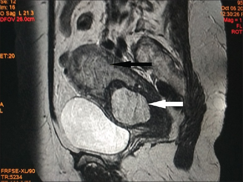
Sagittal T2-weighted magnetic resonance image of the pelvis demonstrating an enlarged uterus with hematometra in the endometrial cavity (black arrow) and a well-defined hyperintense lesion involving the anterior cervix (white arrow)

Sagittal T2-weighted magnetic resonance image of the pelvis demonstrating an enlarged uterus with hematometra in the endometrial cavity (black arrow) and a well-defined hyperintense lesion involving the anterior cervix (white arrow)