
Figure 2.
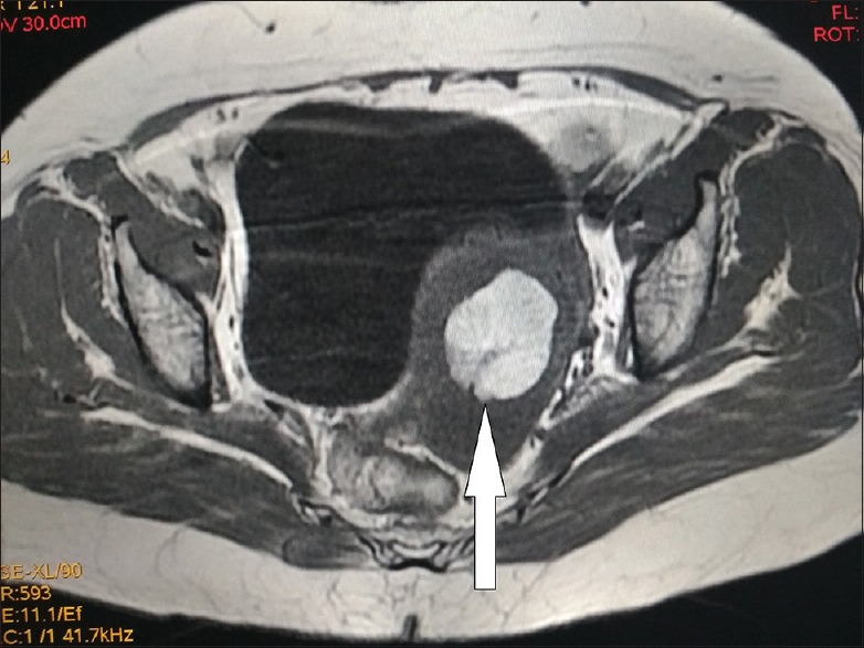
Axial T1-weighted magnetic resonance image of the pelvis demonstrating hyperintense lesion in the cervix representing macroscopic fat-containing lesion proved to be lipoleiomyoma
Official websites use .gov
A
.gov website belongs to an official
government organization in the United States.
Secure .gov websites use HTTPS
A lock (
) or https:// means you've safely
connected to the .gov website. Share sensitive
information only on official, secure websites.
Axial T1-weighted magnetic resonance image of the pelvis demonstrating hyperintense lesion in the cervix representing macroscopic fat-containing lesion proved to be lipoleiomyoma