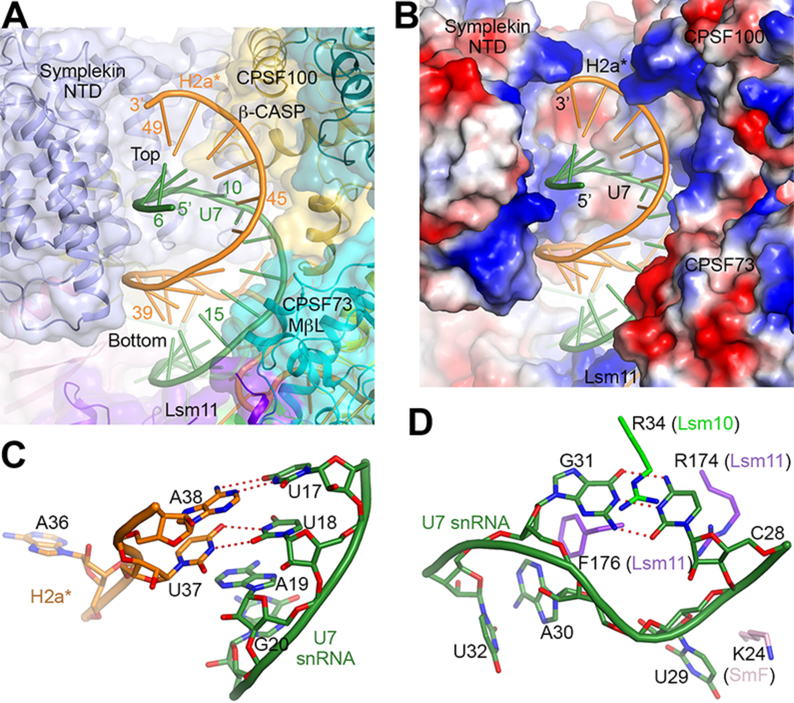
Figure 2. Recognition of the HDE-U7 duplex and the U7 Sm site.
(A) The HDE-U7 duplex is surrounded by CPSF73, CPSF100 and symplekin NTD, shown as a transparent surface. Lsm11 has interactions with the bottom of the duplex. (B) Electrostatic surface of the proteins in the duplex binding site, showing charged interactions with the backbone of the duplex. (C) A U-U base pair at the bottom of the duplex, flanked on the other face by A19 of U7 snRNA. (D) A C-G base pair in the 3′ CUAG sequence of the U7 Sm site. The base pair is flanked on one side by Arg34 of Lsm10, and on the other by Arg174 of Lsm11.

