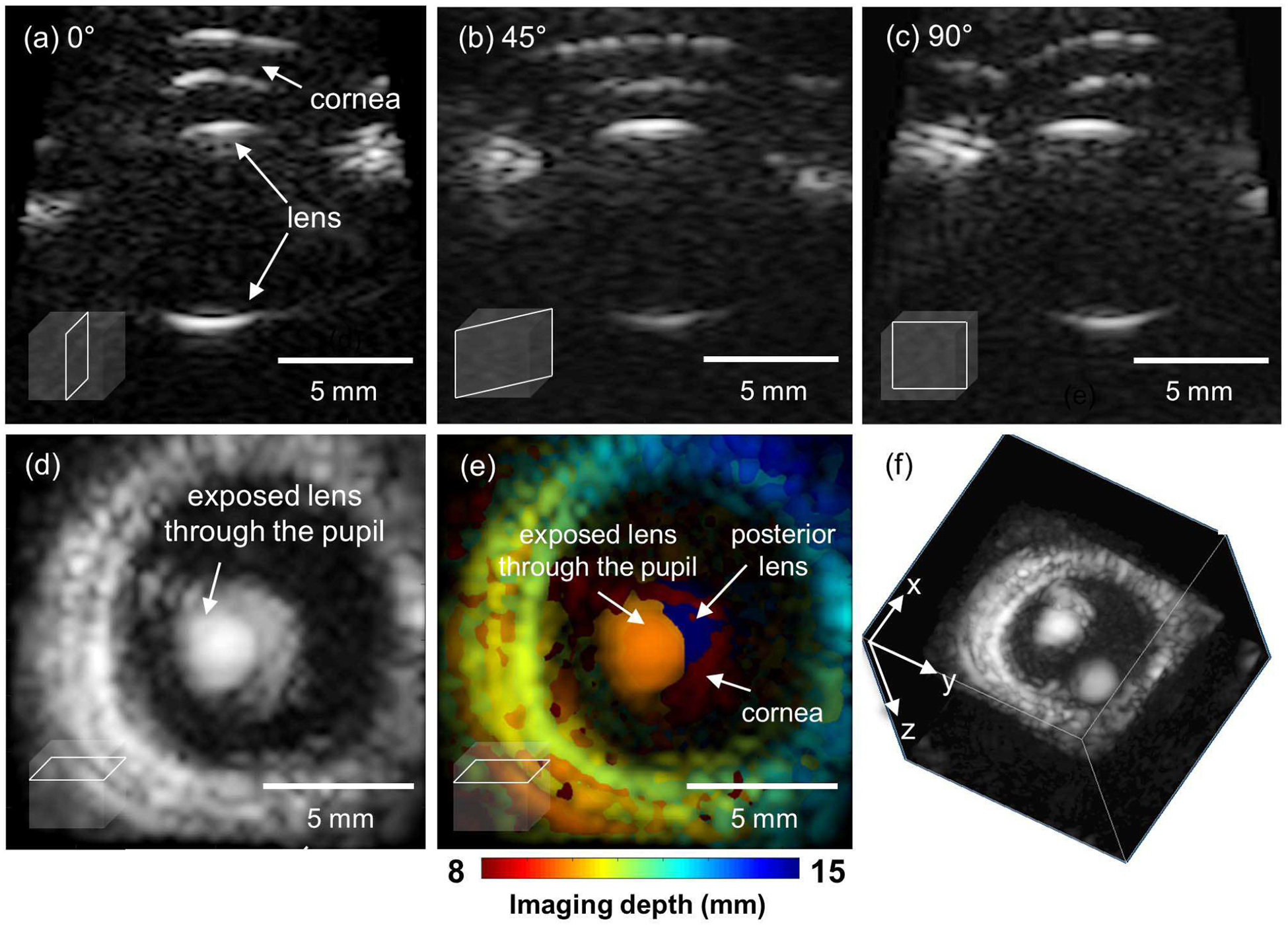
Fig. 7.

Multi-planar imaging of an ex vivo porcine eyeball. (a) The long axis image at azimuthal angle of 0°, (b) 45°, (c) 90°, (d) The image on the coronal plane reconstructed by using the maximum intensity projection. (e) The color-coded depth information superimposed to the coronal image (d). and (f) The 3D volume visualization.
