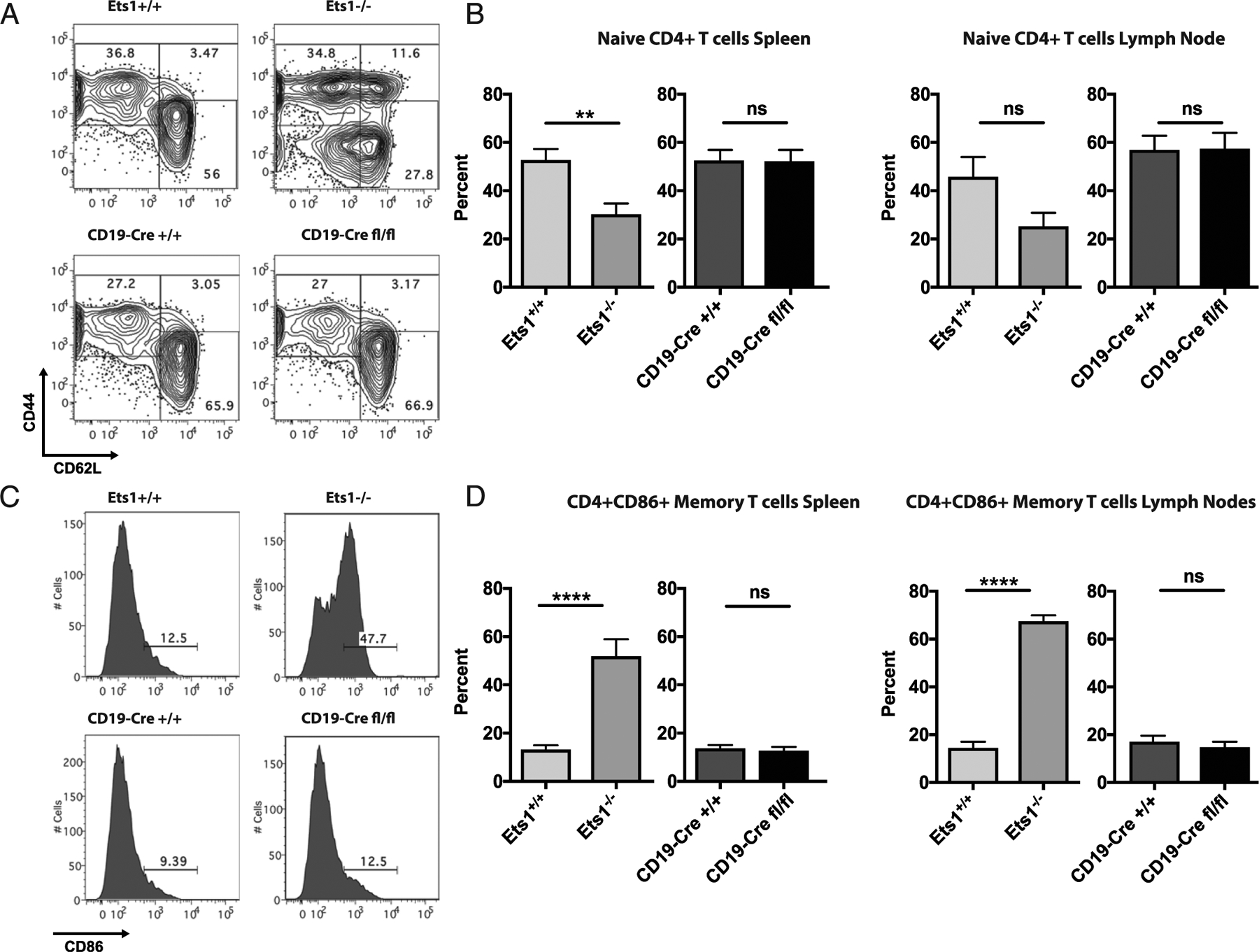
FIGURE 4. Mice with a B cell–specific deletion of Ets1 do not have increased CD4+ T cell activation.

(A) Flow cytometry analysis of CD62L versus CD44 in gated live CD4+ T cells in spleen of the indicated mice. (B) Quantification of the percentages of naive phenotype CD4 T cells (CD62LhiCD44lo) in spleen and lymph nodes of the various strains of mice (n = 7 Ets1+/+, 5 Ets1−/−, 10 CD19-Cre Ets1+/+, and 8 CD19-Cre Ets1fl/fl mice). (C) Flow cytometry analysis of CD86 staining on CD4+ T cells of mice. (D) Quantification of the percent of CD4+ CD86+ T cells in gated live spleen and lymph node cells of the indicated mice (n = 7 Ets1+/+, 5 Ets1−/−, 10 CD19-Cre Ets1+/+, and 8 CD19-Cre Ets1fl/fl mice). **p < 0.01, ****p < 0.0001. ns, not significant.
