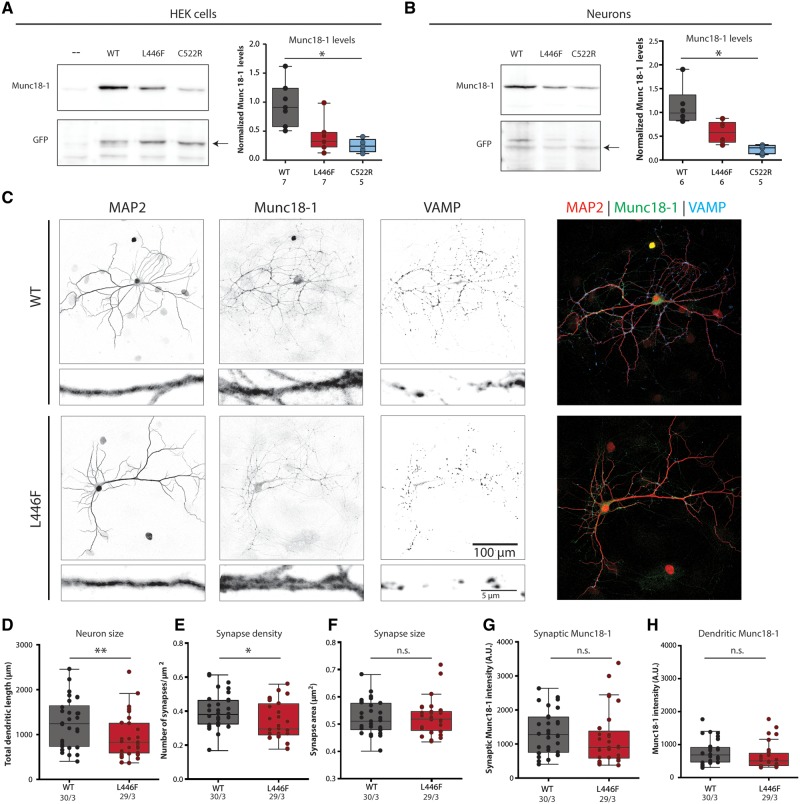
Figure 2.
Cellular stability and morphological characterization of Munc18L446F in Munc18-1 null neurons and HEK293 cells. (A) HEK293 cells were virally infected with Munc18WT, homozygous pathogenic variant Munc18L446F and heterozygous disease variant Munc18C522R. Western blot analysis of normalized Munc18-1 levels shows that Munc18C522R presents significantly lower levels than Munc18WT [Munc18WT median = 0.508, interquartile range (IQR) = 0.340–0.727; Munc18L446F median = 0.187, IQR = 0.134–0.267; Munc18C522R median = 0.143, IQR = 0.085–0.199; P = 0.0006, Kruskal-Wallis test with post hoc Dunn’s multiple comparisons test]. Munc18L446F has no significant changes in levels compared to either Munc18WT and disease variant Munc18C522R. Munc18 levels were normalized to GFP levels. Relative Munc18 levels were normalized to the mean Munc18WT levels for visualization. (B) Munc18WT, homozygous disease variant Munc18L446F and heterozygous disease variant Munc18C522R were expressed in Munc18-1 null neurons through lentiviral infection. Protein levels of Munc18C522R are lower than Munc18WT (Munc18WT median = 1.587, IQR = 1.401–2.278; Munc18L446F median = 0.978, IQR = 0.578–1.296; Munc18C522R median = 0.397, IQR = 0.228–0.526; P = 0.0012, Kruskal-Wallis test with post hoc Dunn’s multiple comparisons test), whereas levels of Munc18L446F are not significantly different from Munc18WT and Munc18C522R. Munc18 levels were normalized to GFP levels. Relative Munc18 levels were normalized to the mean Munc18WT levels for visualization. (C) Representative images (with zoom) of Munc18-1 null neurons expressing Munc18WT or Munc18L446F, stained for MAP2 (dendritic marker), Munc18-1 and VAMP (synaptic marker). (D) Total dendritic length is decreased in Munc18L446F neurons (Munc18WT median = 1243, IQR = 738–1645; Munc18L446F median = 833.4, IQR = 592.5–1254; P = 0.035, Mann-Whitney U-test). (E) Munc18L446F neurons show decreased number of synapses per μm2 dendrite (Munc18WT median = 0.382, IQR = 0.326–0.465; Munc18L446F median = 0.296, IQR = 0.262–0.444; P = 0.028, unpaired t-test). (F) Average synapse area is not altered between neurons expressing Munc18WT or Munc18L446F (Munc18WT median = 0.511, IQR = 0.480–0.577; Munc18L446F median = 0.519, IQR = 0.477–0.547; P = 0.894, Mann-Whitney U-test). (G and H) Munc18L446F neurons do not present lower Munc18-1 levels in synapses (G) (Munc18WT median = 1276, IQR = 754.1–1789; Munc18L446F median = 893.8, IQR = 584–1376; P = 0.091, Mann-Whitney U-test) or in dendrites (H) (Munc18WT median = 654.6, IQR = 471–894; Munc18L446F median = 479.7, IQR = 353.6–641.7; P = 0.094, Mann-Whitney U-test) by immunocytochemistry. The number of analysed cells and number of independent cultures tested is indicated below the graphs. *P < 0.05, **P < 0.01.

