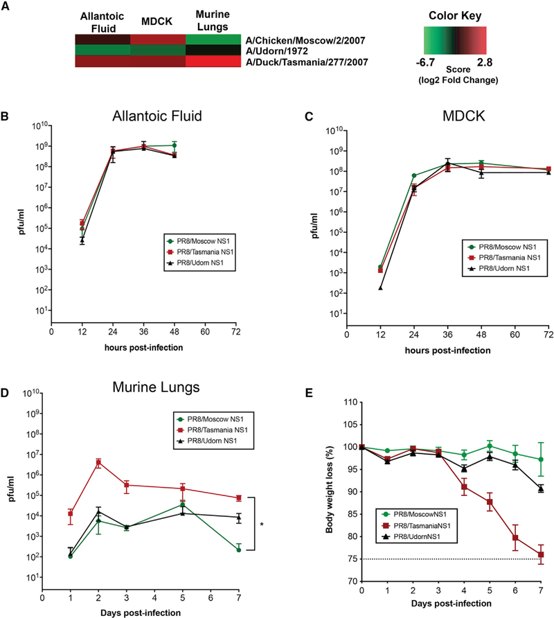
Figure 3. Viral Library Profile Dynamics Can Be Reproduced in Single-Virus Experiments in a A/Puerto Rico/8/1934 (H1N1) Background In Vivo.
(A) Three representative viruses showing different viral trends in the library were selected based on differences in barcode abundance.
(B–D) Single-virus infections were conducted in triplicate using 10-day-old embryonated chicken eggs (B), MDCK cells (C), and 8-week-old C57BL/6 mice (D). Viral replication was quantified at different time points post-infection.
(E) Body weight loss of infected mice was monitored daily.
Error bars depict the SD. *p < 0.05.

