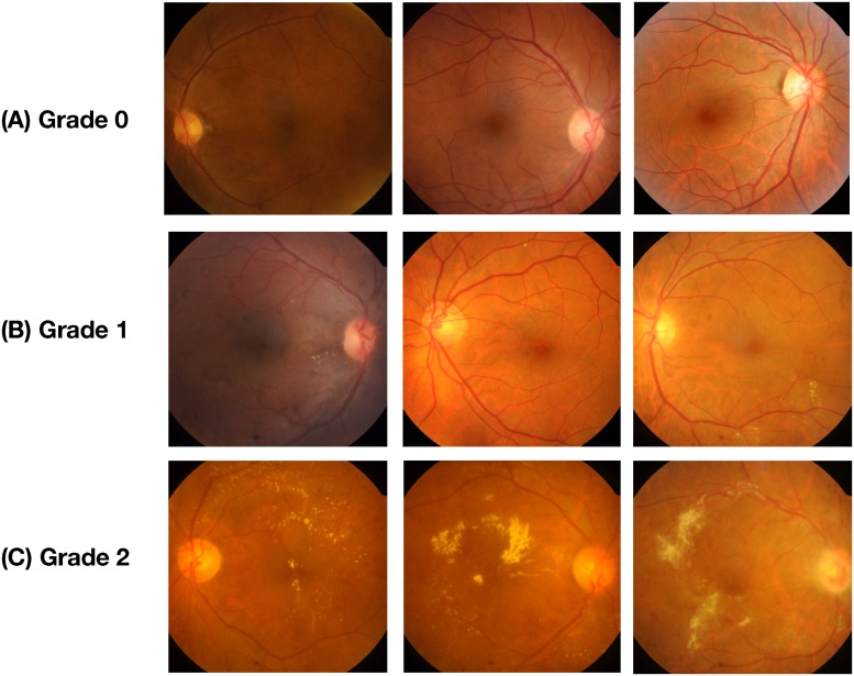
Fig 1. Representative fundus images with different pathological severity of DME.
(A) Denotes the images of Grade 0 severity which have no hard exudates, (B) Represents Grade 1 severity which has exudates outside the radius of one disc diameter from the macula center, and (C) Denotes the fundus under the Grade 2 severity category, with exudates within the radius of one disc diameter from the macula center.

