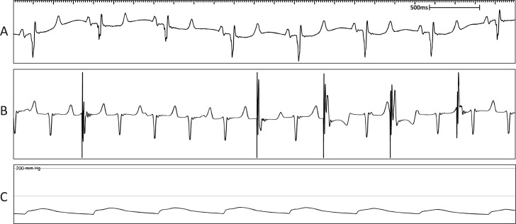
Fig 7. Representative surface ECG recordings following creation of AVB in sheep M9 showing a fast, fascicular rhythm.
(A) Surface ECG depicting normal sinus rhythm. (B) Surface ECG showing AVB evident by AV dissociation with intermittent pacing due to the presence of a fast, fascicular rhythm competing with the pacemaker programmed at 80 bpm. (C) Mean arterial blood pressure trace indicating maintenance of blood pressure during the procedure.

