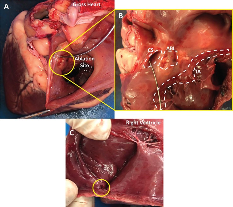
Fig 11. Gross anatomy of an explanted sheep heart showing landmarks, ablation lesions and lead attachment site.
(A) This view depicts the gross heart anatomy with the view looking into the right atrium. The ablation site is shown within the circle. A wire was placed in the coronary sinus for orientation purposes. (B) Zoomed in perpendicular view of the coronary sinus (CS), tricuspid annulus (TA) and the ablation lesions (ABL). (C) View showing the lead attachment site within the circle at the apex of right ventricle.

