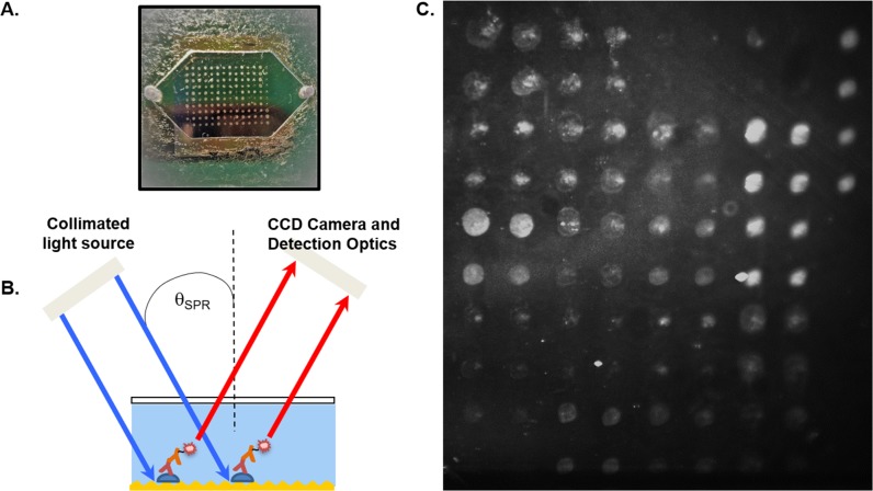
Fig 1. The GC-FP platform and its linear range of IgG antibody detection.
(A) After being coated with antigens, a GC-FP biochip is assembled with a gasket and window to form a microfluidic chamber, where serum samples and other reagents can be applied. (B) GC-FP analysis on the gold-coated biochip involves using a fluorophore-labelled secondary antibody that couples with the surface plasmon field to emit enhanced fluorescent signal. (C) A representative GC-FP image is shown, containing various LD targets. The fluorescence intensity at each spot ROI corresponds to the amount of detected antibody.

