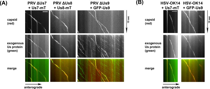
Fig 5.
(A) TIRF microscopy of live SCG axons grown in compartmentalized cultures. Cells were transduced with indicated PRV proteins, infected with red capsid tagged PRV mutant lacking the corresponding protein and imaged at ~19frames/s at 12-14hpi. Axonal co-transport of the viral envelope protein was observed with all viral capsids. (B) Same as in (A), but with transduction of HSV-1 proteins and red capsid tagged HSV-1 wild-type virus.

