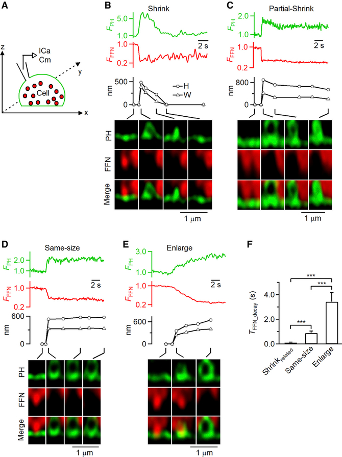
Figure 2. Observation of Ω-Shaped Membrane Profile Shrinking and Enlargement while Releasing FFN511.
(A) Setup diagram. Vesicles were loaded with FFN511 (red, pseudo-color), the PM was labeled with PHG (green), and ICa and Cm were recorded via a pipette.
(B–E) FPH, FFN511 fluorescence (FFFN; normalized to baseline), PH-Ω height (H; circles) and width (W; triangles), and sampled images at times indicated with lines showing release of FFN511 for shrink fusion (B), partial shrink fusion (C), same-size fusion (D), and enlarge fusion (E).
(F) 20%–80% decay time of FFN511 (TFFN_decay, mean + s.e.m.) for shrink fusion (8 events), partial shrink fusion (13 events), same-size fusion (53 events), and enlarge-fusion (18 events). Data are from 71 cells under STED xz/yfix imaging of PHG/FFN511. ***p < 0.001; ANOVA test.

