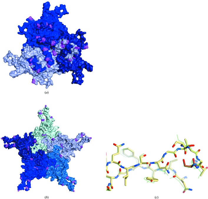
Figure 4.
AAVhu.37 VP3 monomers shown in shades of blue, highlighting amino-acid differences (pink) between AAVhu.37 and AAVrh.10 relative to the (a) threefold and (b) fivefold axes of symmetry. (c) Comparison of the VR-I loop of AAV8 (shown in green as lines) and AAVhu.37 (shown as sticks with C atoms colored yellow; amino acids 262–275). Residue 269, the single amino-acid difference in VR-I between the two serotypes, is indicated by darker coloring (orange C atoms).

