. 2020 Jan 23;9:e52572. doi: 10.7554/eLife.52572

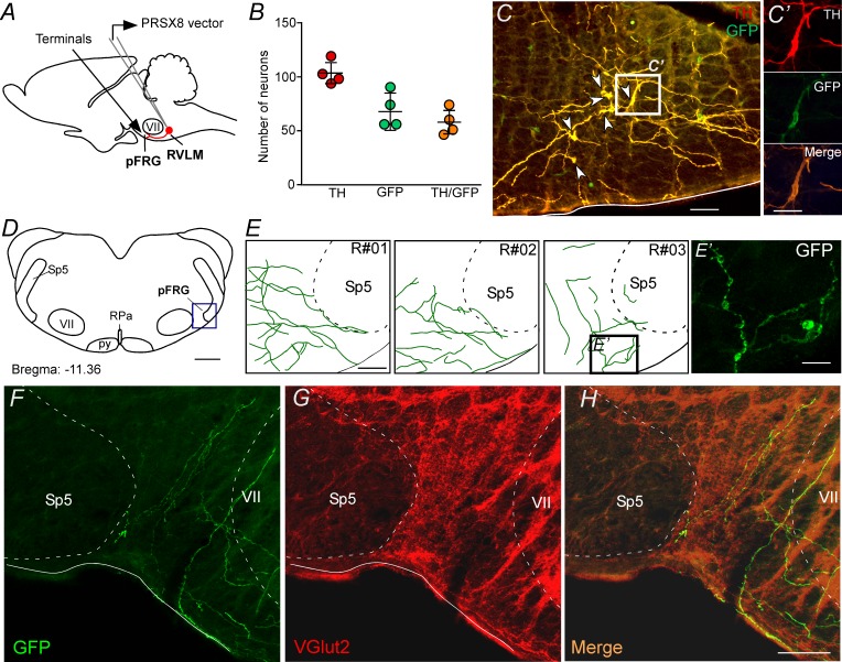
Figure 3. pFRG receives glutamatergic inputs from catecholaminergic C1 neurons.

(A) Experimental design. (B) The number of TH and GFP neurons in the RVLM (N = 4). Cell count was obtained in six coronary brain sections (40 μm with 240 μm intervals between slices) from each rat. (C, C’) Single case showing that the C1 neurons are transfected by the lentivirus PRSX8-ChR2-eYFP. (D, E) Schematic drawing of a coronal brain section showing the distribution of GFP terminals in the pFRG region in three rats (bregma level: −11.36 mm in accordance with Paxinos and Watson, 2007). (E’) Photomicrography showing terminals into the pFRG region of the R#03. (F–H) Photomicrography showing fibers and terminals expressing lentivirus (GFP, green) and glutamate (VGlut2, red) in the pFRG region. Abbreviations: Fn, facial nerve; GFP, green fluorescent protein; pFRG, parafacial respiratory group; NTS, nucleus of the solitary tract; py, pyramid tract; RPa, raphe pallidus; RVLM, rostral ventrolateral medulla; Sp5, spinal trigeminal tract; TH, tyrosine hydroxylase; VGlut2, vesicular glutamate transporter 2; VII, facial motor nucleus. Scale bars in panels (C) and (E–H) = 50 μm, (C’), (E’) and (H’) = 20 μm, (D) = 1 mm.

Figure 3.

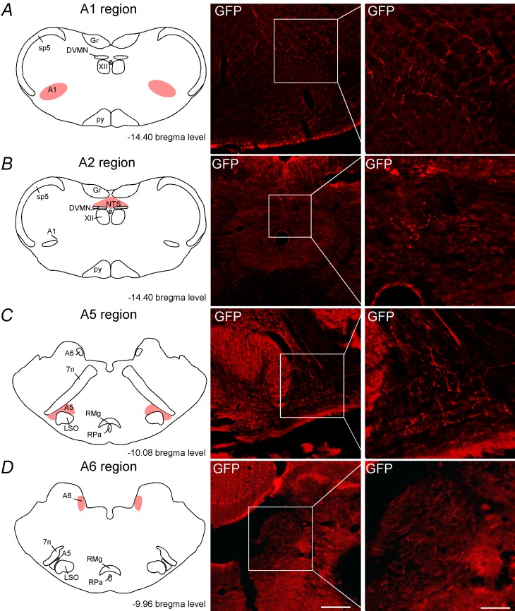
Figure 3—figure supplement 1. C1 neurons project to the noradrenergic neurons.

Figure 3—figure supplement 1.

Schematic drawing showing the location of the catecholaminergic (A) A1 region, (B) A2 region, (C) A5 region and (D) A6 region. The photomicrographs show fibers and terminals arising from C1 neurons. Abbreviations: DVMN, dorsal vagus motor nucleus; Fn, facial nerve; Gr, gracile nucleus; LSO, lateral superior olive; NTS, nucleus of the solitary tract; py, pyramid tract; RMg, raphe magnus; RPa, raphe pallidus; Sp5, spinal trigeminal tract; XII, hypoglossal nucleus. Scale bar = 100 μm.
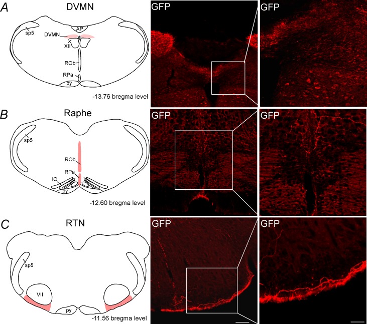
Figure 3—figure supplement 2. C1 neurons project to DVMN, medullary aphe and RTN regions.

Figure 3—figure supplement 2.

Schematic drawing showing the location of the (A) dorsal vagus motor nucleus (DVMN), (B) the medullary raphe and (C) the retrotrapezoid nucleus (RTN). The photomicrographs show fibers and terminals arising from C1 neurons. Abbreviations: AP, area postrema; DVMN, dorsal vagus motor nucleus; IO, inferior olive; py, pyramid tract; ROb, raphe obscurus; RPa, raphe pallidus; Sp5, spinal trigeminal tract; XII, hypoglossal nucleus. Scale bar = 100 μm.