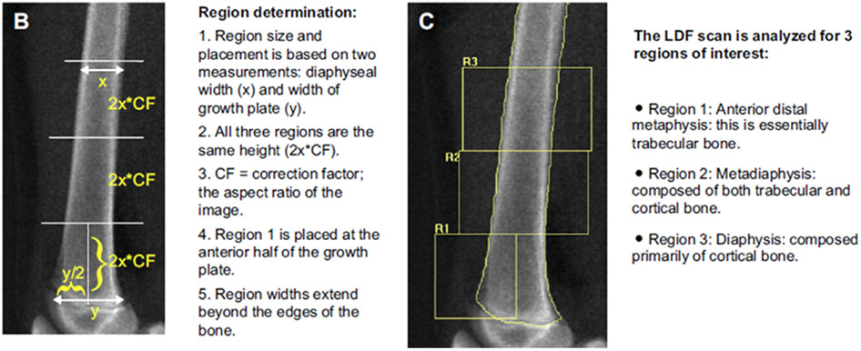
Fig. 1.
Lateral distal femur scan showing the 3 regions of interest. Region (R)1 is metaphyseal and is comprised primarily of trabecular bone, R2 is metadiaphyseal and comprised of a mixture of cortical and trabecular bone, and R3 is diaphyseal which is primarily cortical bone. Reproduced from Zemel et al, (97).

