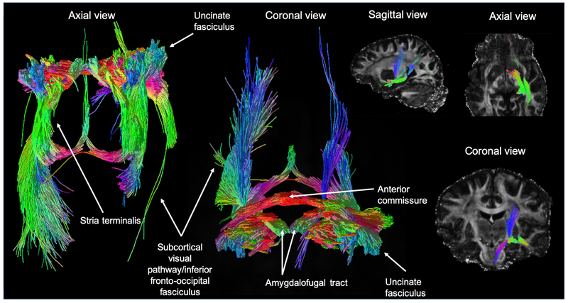
Fig. 1.
An axial, coronal and sagittal view of probabilistic tractography seeded at all amygdala nuclei combined. White matter tracts reconstructed include: the stria terminalis, the uncinate fasciculus, a subcortical visual pathway passing along the inferior fronto-occipital fasciculus, the amygdalofugal tract and the anterior commissure. The color of streamlines is determined by their directionality.

