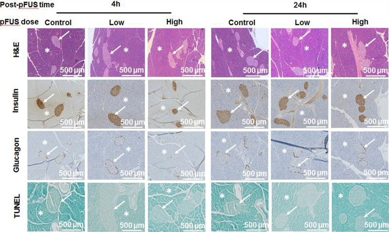
Figure 2. Histological analysis of pFUS treated pancreases:

Samples of pFUS treated pancreases stained with H&E, insulin, glucagon and TUNEL. Asterisks show exocrine and arrows show endocrine (i.e. islets) components of the pancreas.

Samples of pFUS treated pancreases stained with H&E, insulin, glucagon and TUNEL. Asterisks show exocrine and arrows show endocrine (i.e. islets) components of the pancreas.