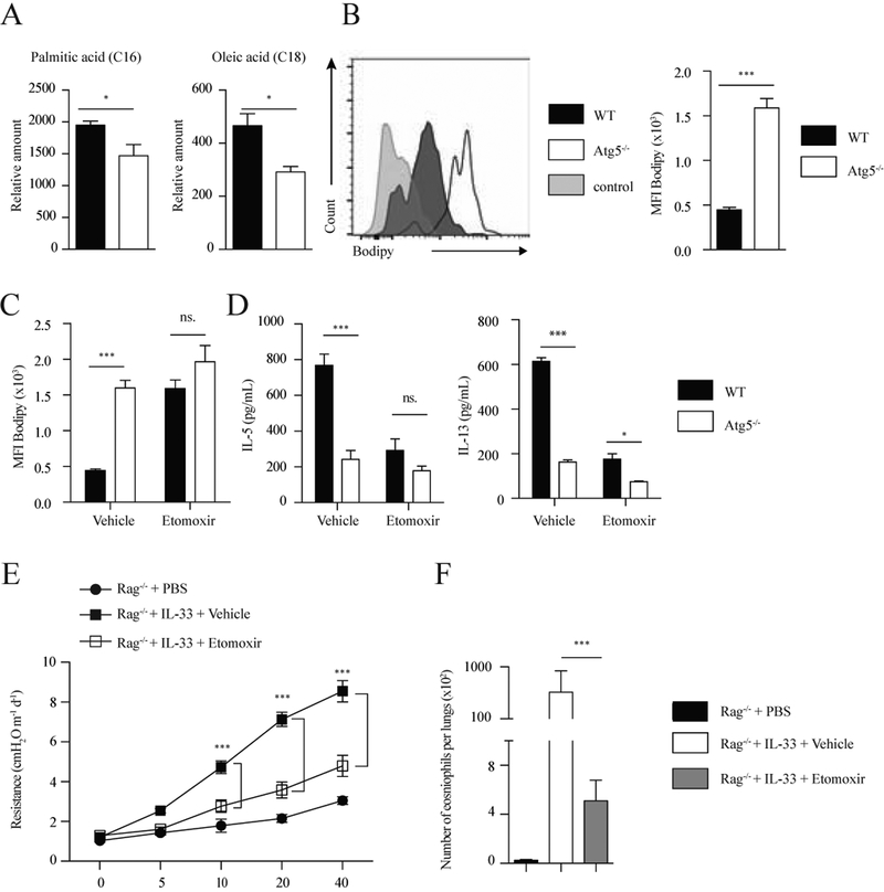
Figure 5: Lack of autophagy inhibits lipolysis in activated ILC2s.
(A) Free fatty acid abundance in ILC2s isolated from WT control and Atg5−/− mice quantified by GCxGC/MS. Data represent 4 biological replicates per genotype. (B) Bodipy histogram (left) and quantification as MFI (right) in WT control and Atg5−/− ILC2s from lungs treated with rmIL-33 (10ng/mL) for 24 hours. The level of stain control is shown as a gray-filled histogram. (C) Bodipy quantification as MFI in WT control and Atg5−/− ILC2s from lungs were treated with rmIL-33 (10ng/mL) and stimulated with vehicle or etomoxir (20ng/μL) for 24 hours. WT control and Atg5−/− ILC2s from lungs were treated with rmIL-33 (10ng/mL) and stimulated with vehicle or etomoxir (20ng/μL) for 24 hours. (D) The levels of IL-5 (left) and IL-13 (right) were measured by ELISA on the culture supernatants, n=6. A cohort of Rag−/− mice was intranasally treated with PBS or rmIL-33 (0.5μg) on days 2–4. Mice were i.p. injected with etomoxir (15mg/kg) or vehicle on days 1 and 3. Measurement of lung function and BALF analysis followed on day 5. (E) Lung resistance. (F) Total number of eosinophils in BALF. Data are representative of at least three independent experiments, n=6. Error bars are the mean ± SEM. Student’s t-test, *p < 0.05, **p < 0.01, ***p < 0.001.

