Figure 1.

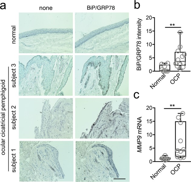
ER stress is elevated in ocular cicatricial pemphigoid. (a) BiP/GRP78 staining was analyzed in conjunctival biopsies of three patients with ocular cicatricial pemphigoid (OCP) and three normal subjects by immunohistochemistry. A representative image is shown for normal tissue. Consecutive sections lacking the primary antibody were used as negative controls. Scale bar, 100 μm. (b) BiP/GRP78 staining intensity in conjunctival epithelium was measured in at least two different histological areas for each subject using ImageJ. (c) Conjunctival epithelium was collected by impression cytology from nine patients with OCP and six normal subjects. The expression of MMP9 was determined by qPCR. The box and whisker plots show the 25 and 75 percentiles (box), the median, and the minimum and maximum data values (whiskers). Significance was determined using Mann-Whitney test. **p < 0.01.
