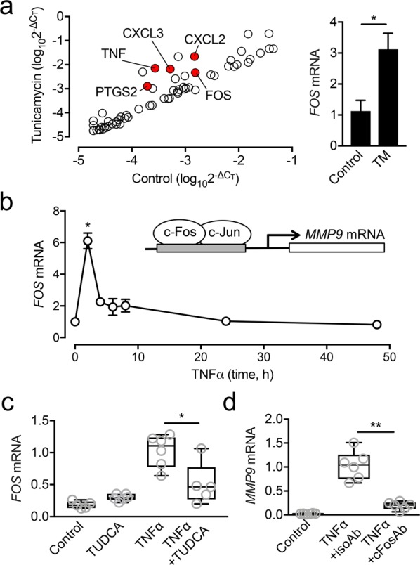
Figure 5.

ER stress induces MMP9 expression by promoting FOS transcription. (a) Monolayer cultures of human corneal epithelial cells were incubated with 10 µg/ml tunicamycin for 2 h. The relative expression of genes involved in the inflammatory response was evaluated with a human inflammatory response and autoimmunity PCR array. The red dots in the scatter diagram indicate at least 2-fold significant upregulation compared to control. No gene was significantly downregulated. (b) Multilayered cultures of human corneal epithelial cells were incubated with 40 ng/ml TNFα at different time points. The expression of FOS was analyzed by qPCR. (c) The effect of TUDCA on FOS expression was measured by qPCR following 2 h incubation with TNFα. (d) The effect of a function-blocking antibody to human c-Fos (cFosAb) on MMP9 expression was measured by qPCR following 48 h incubation with TNFα. An isotype-matched antibody (isoAb) served as control. Results in (a,b) represent three independent experiments. Results in (c,d) represent two independent experiments performed in triplicate. Data in (a,b) represent the mean ± SEM. The box and whisker plots show the 25 and 75 percentiles (box), the median, and the minimum and maximum data values (whiskers). Significance was determined using Student’s t test (a), Kruskal-Wallis with Dunn’s post hoc test (b) and Mann-Whitney test (c,d). *p < 0.05; **p < 0.01. TM, tunicamycin.
