Figure 5.
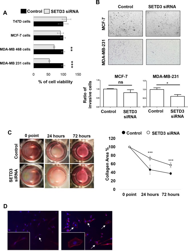
The inhibition of SETD3 expression blocks the viability and invasive phenotype of MDA-MB-231 breast cancer cells. (A) MTT assay reveals a significant effect of SETD3 on cell viability in the aggressive basal cell lines MDA-MB-231 and MDA-MB-468. (B) After 48 hours of culture with siRNA to inhibit the expression of SETD3, a Matrigel transwell invasion was performed. MCF-7 cells are poorly invasive luminal cells while MDA-MB-231 cells are highly invasive basal cells. (C) Collagen contraction, and (D) immunofluorescence assays were performed in MDA-MB-231 cells. Upper (B) and left (C) panels show representative images of invasion and collagen contraction respectively, and the ratio of invasive cells or the percent of contraction collagen area were graphed. Data represent the mean ± SEM from 3 independent experiments in triplicates. Bars or points with asterisks represent comparisons with statistically significant differences (P < 0.05). For immunofluorescence (D) representative images are shown. Actin filaments were stained with phalloidin (red) and nuclei with 4′,6′-diamidino-2-phenylindole (DAPI, blue). White arrows represent the normal cells (left panel) and those that have modified their morphology after the depletion of SETD3 (right panel). Magnification 200×.

