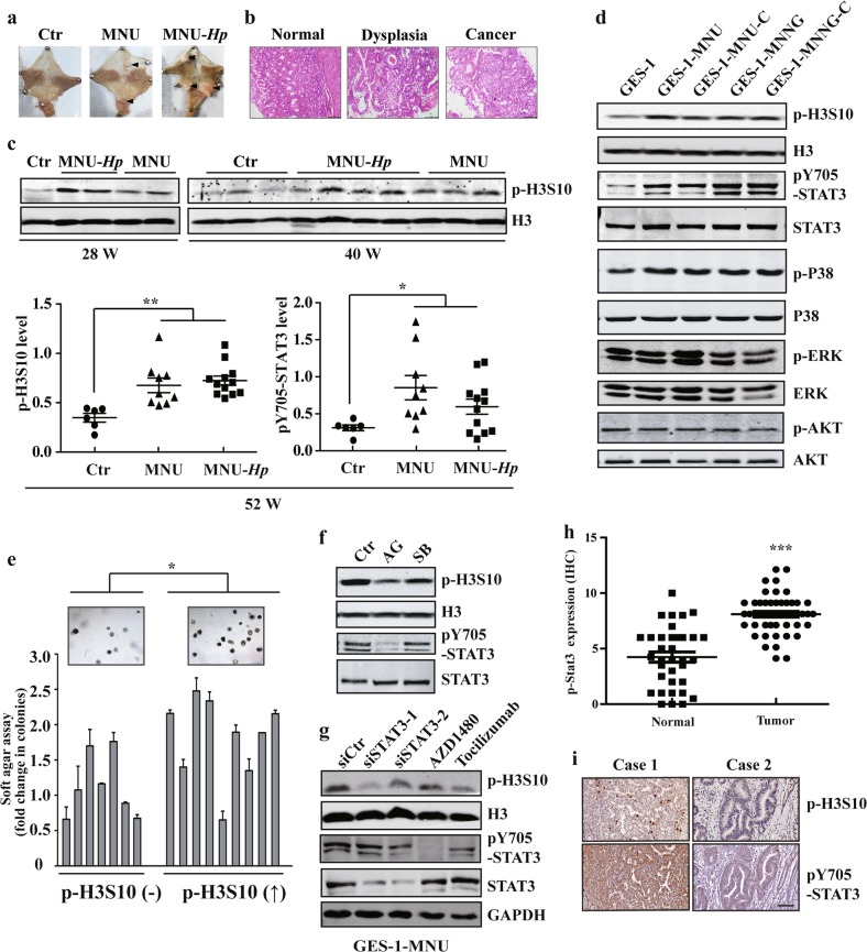
Fig. 1. STAT3 contributes to enhanced H3S10 phosphorylation in gastric carcinogenesis.
a Representative image of NOC (N-methyl-N-nitroso-urea, MNU) and H. pylori plus NOC -induced mouse model of gastric cancer. b HE staining of stomach tissues from the carcinogen-treated mice. c Histone H3 phosphorylation was analyzed by WB of stomach tissues from the carcinogen-treated mice at different times. d WB analysis of signaling pathways related to cell proliferation in GES-1 and NOC-treated cells with the indicated antibodies. e Cell colony-forming ability in soft agar of p-H3S10 increased or unchanged subcloned NOC-treated cells. f, g AG490 50 μM, SB203580 10 μM, AZD1480 2 μM, Tocilizumab 10 μM, scramble siRNA (siCtr) or STAT3 siRNA were used to treat the MNU-transformed cells, respectively. The level of p-H3S10 was determined by WB. h STAT3 expression score by IHC in 52 paired gastric cancer tissues. i The representative images of STAT3 and p-H3S10 levels in human gastric cancer tissues by IHC. Scale bar: 200μm. The analyses were repeated three times, and the results were expressed as mean ± SD. */#p < 0.05, **p < 0.01, ***p < 0.001. GES-1-MNNG or GES-1-MNU: MNNG- or MNU-induced malignantly transformed GES-1 cell; GES-1-MNNG-C or GES-1-MNU-C: subcloned cells derived from GES-1-MNNG or GES-1-MNU.

