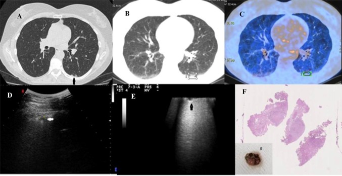
Fig. 5.
a–c18F-FDG PET/CT combined image in patient with history of endometrioid carcinoma and tobacco habits showing low increased glucose uptake and glycolysis on the right lung and calcified micro-nodule in the posterior basal segment of the left lower lobe (black arrow in a); d TUS image of the same patient (convex probe: 5 MHz) showing mixed hypoechoic micro-nodule in the posterior basal segment of the left lung (white arrow); e VATS-US (intraoperative linear probe) showing a hypoechoic structure and well-delimited margins of the left micro-nodule (black arrow); f histological examination of the micro-nodules of the left lower lobe excised during VATS (specimen showed in box G) showing a final diagnosis of histiocytosis X

