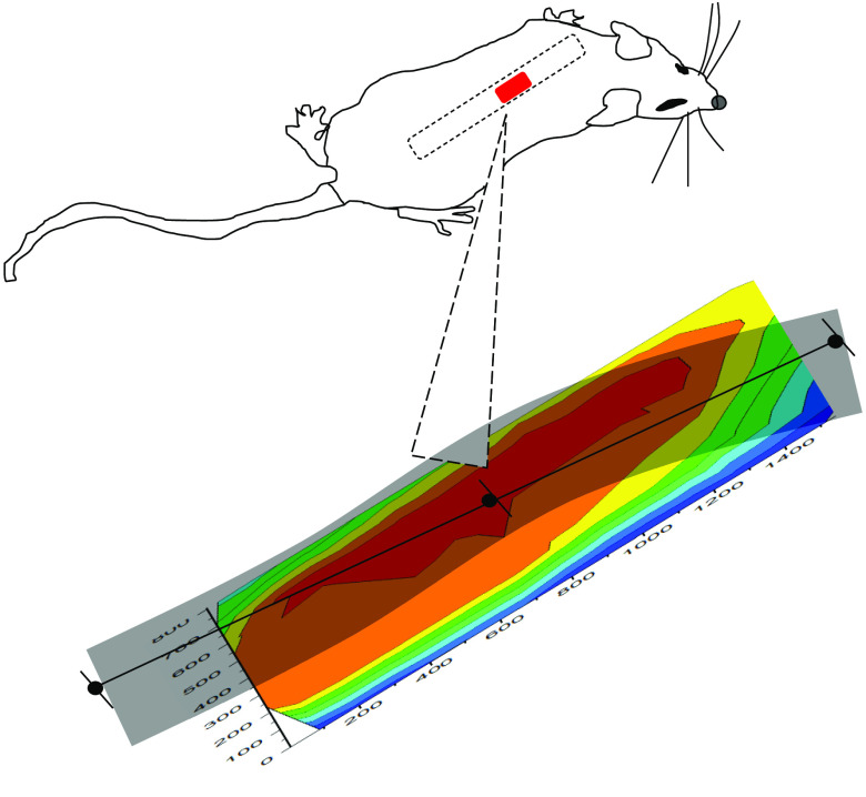
Fig. 10.
Image of surgical field including spinal cord based on Hct values obtained in a scan trajectory [Fig. 4(b)] designed so that no point is probed twice so as to avoid photobleaching effects from an early test scan causing systematic artifacts in later images. Note the systematic fall off of the apparent Hct on the edges of the image due at least in part because of topographic variation, i.e., curvature. The spatial scale is indicated on both axes using the time from the beginning of the first scan in seconds since the actual distances are as shown in Figs. 4(a) and 4(b), and the possibility of photobleaching artifacts is determined by the time duration of the laser exposure.

