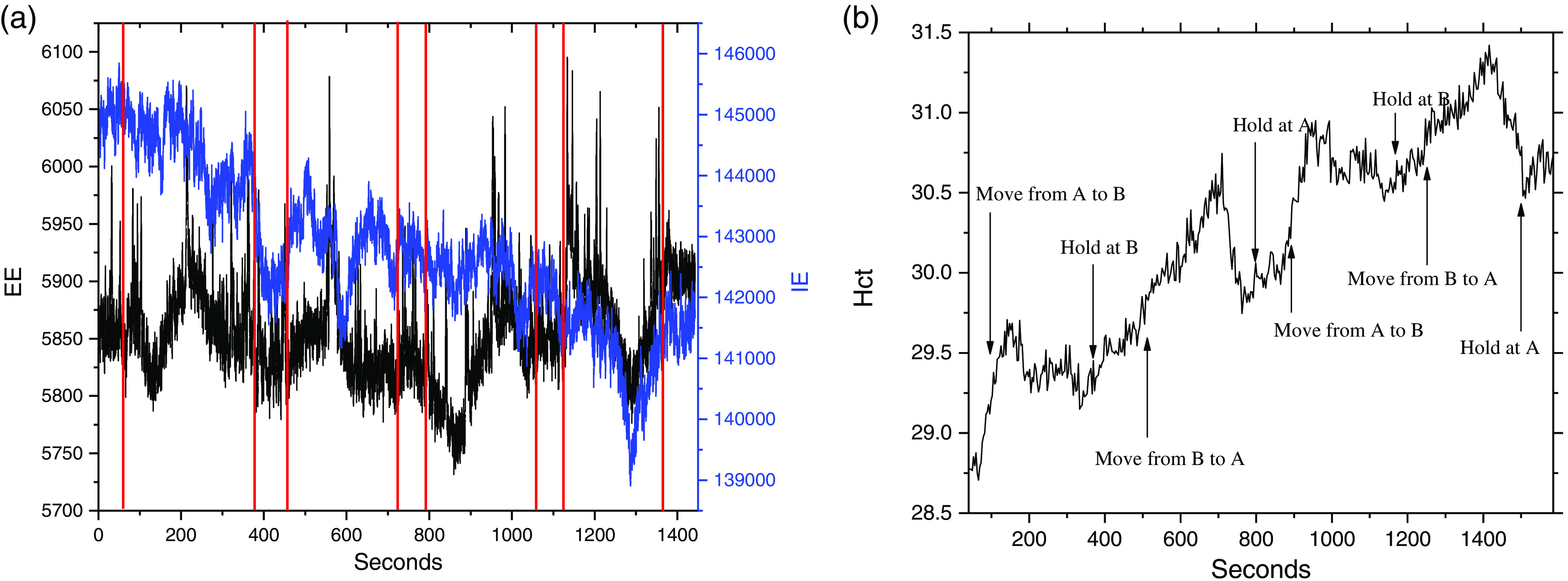
Fig. 6.

(a) The EE and IE obtained while scanning a cord for the purpose of showing how this imaging works. This scan did not follow the protocol described in Sec. 2. In this case, the scan started at A with the rat motionless for 80 s, then scanned to B in 200 s, stayed at B for 80 s, scanned back to A in 200 s, stayed at A for 80 s, rescanned from A to B in 200 s, stayed at B for 80 s, rescanned back to A in 200 s, and collected last 80 s at A. The laser probing was at the location indicated in (b): (1) at beginning and end of experiment and (2) between the red lines in (a).
