Fig. 6.
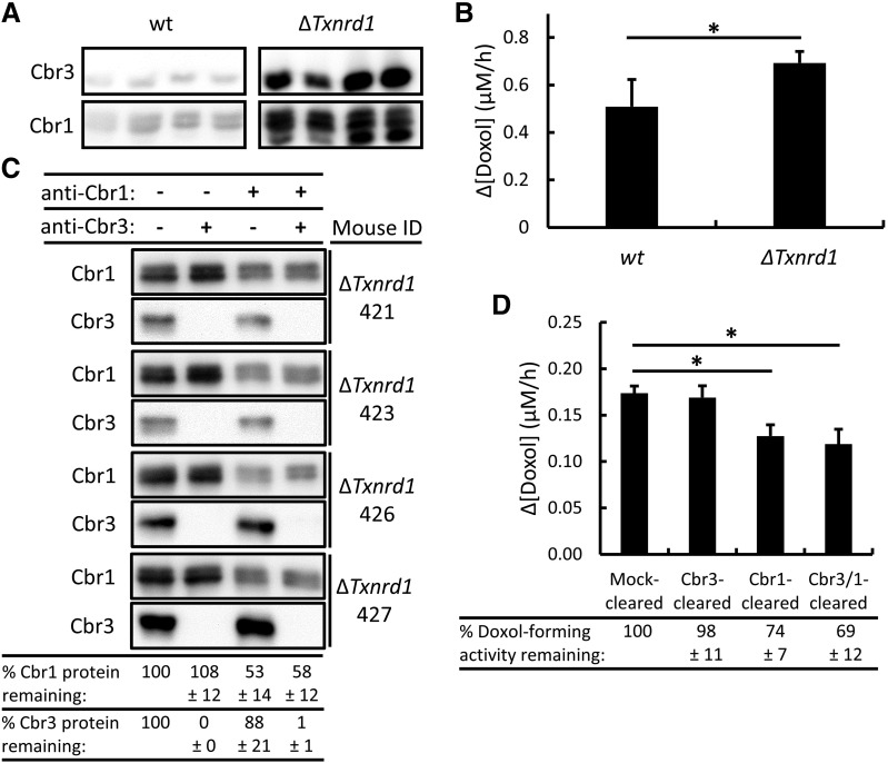
Immunoclearing of Cbr1 and/or Cbr3 and its effect on Doxol-forming activity in ΔTxnrd1 liver cytosols that overexpress Cbr3. (A) Immunoblot analysis of Cbr3 and Cbr1 in wild-type (wt) and ΔTxnrd1 mouse liver cytosols; 18 µg of total cytosolic protein loaded per lane. The top panel shows a blot probed with Cbr3-specific antibody. The bottom panel shows the same blot reprobed with Cbr1-specific antibody; because the blot was not stripped, the fast-migrating Cbr3 signal remained visible. (B) NADPH-dependent Doxol formation by wild-type and ΔTxnrd1 cytosols. Reaction conditions were 200 µM NADPH, 200 µM Dox, and 12-µg equivalents of cytosolic protein; incubations were for 1 hour at 37°C. Doxol levels were measured by LC-MS/MS. (C) Immunoblot analysis of immunocleared ΔTxnrd1 cytosols. Cbr1 and Cbr3 protein levels were determined by densitometry, and the % protein remaining after immunoclearing, relative to mock-cleared samples, is shown at bottom. (D) NADPH-dependent Doxol formation by mock-cleared, Cbr3-cleared, Cbr1-cleared, and Cbr3- and Cbr1-cleared ΔTxnrd1 cytosols. Reaction conditions and incubations were as described in (B), except 50 µM Dox was used. Doxol levels were measured by LC-MS/MS. The % Doxol-forming activity remaining after immunoclearing, relative to mock-cleared samples, is shown at bottom. Error bars represent 1 S.D.; n = 4 for all reactions; *P < 0.05 by Student’s t test.

