
Figure 1.
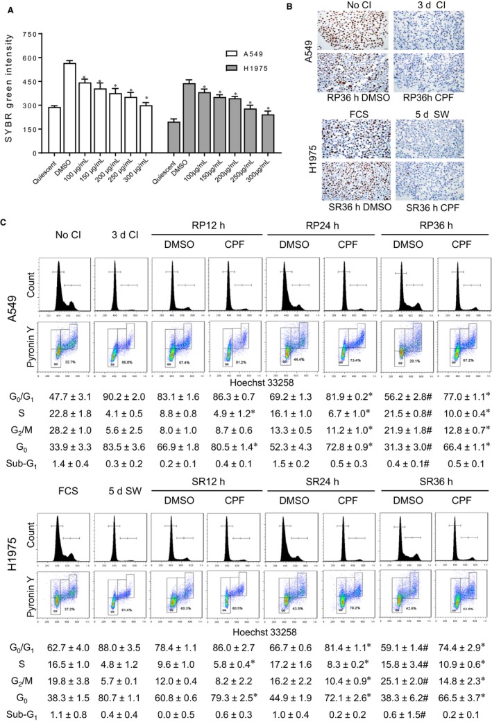
CPF suppresses proliferative switch from G0 state in lung cancer cells. A, CPF or DMSO was administered upon releasing from quiescence for 36 h. The treated two lung cancer cell lines together with the quiescent baseline cells were incubated with lysis buffer containing SYBR Green, and the DNA contents were measured. *P < .05 vs DMSO. B, The non‐quiescent and quiescent cells and the cells treated with DMSO or CPF at GI90 upon cell cycle re‐entry for 36 h were harvested for analysis of Ki‐67 by immunocytochemical staining. No CI: no contact inhibition; 3d CI: contact inhibition for 3 d; RP36h DMSO: treatment of replated cells with DMSO; RP36h CPF: treatment of replated cells with CPF at GI90. FCS: no removal of foetal calf serum; 5d SW: serum withdrawal for 5 d; SR36h DMSO: treatment of serum‐replenished cells with DMSO; SR36h CPF: treatment of serum‐replenished cells with CPF at GI90. C, The non‐quiescent cells, quiescent cells and cells after releasing from G0 at indicated time were stained with Hoechst 33258 and Pyronin Y. Representative images and quantification data after analysis of Hoechst 33258 alone or both Hoechst 33258 and Pyronin Y are shown. Data are expressed as the mean ± SD from three experiments. *P < .05 vs DMSO at each time‐point. #P > .05 vs No CI or FCS