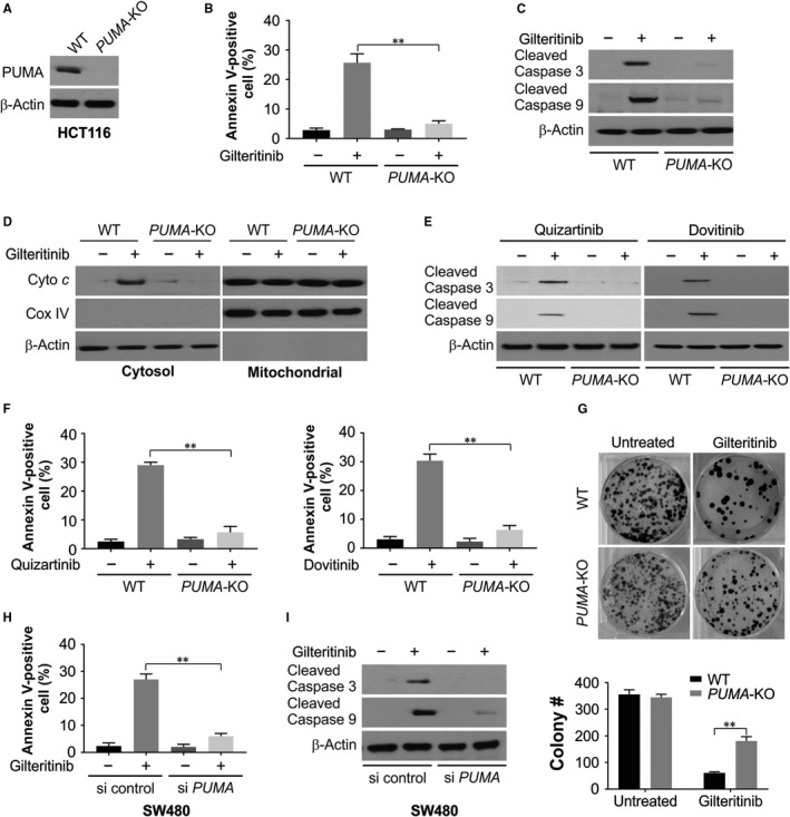
Figure 3.

PUMA is required for the apoptotic and antitumour effects of gilteritinib via mitochondrial pathway. A, PUMA expression was analysed by Western blotting in WT and PUMA‐KO HCT116 cells. B, WT and PUMA‐KO HCT116 cells were treated with 50 nmol/L gilteritinib for 24 h. Apoptosis was analysed by Annexin V/PI staining followed by flow cytometry. C, WT and PUMA‐KO HCT116 cells were treated with 50 nmol/L gilteritinib for 24 h. Cleaved caspase 3 and 9 were analysed by Western blotting. D, Cytosolic fractions isolated from WT and PUMA‐KO HCT116 cells treated with 50 nmol/L gilteritinib for 24 h were probed for cytochrome c by Western blotting. β‐Actin and cytochrome oxidase subunit IV (Cox IV), which are expressed in cytoplasm and mitochondria, respectively, were analysed as the control for loading and fractionation. E, WT and PUMA‐KO HCT116 cells were treated with 50 nmol/L quizartinib or dovitinib for 24 h. Cleaved caspase 3 and 9 were analysed by Western blotting. F, WT and PUMA‐KO HCT116 cells were treated with 50 nmol/L quizartinib or dovitinib for 24 h. Apoptosis was analysed by Annexin V/PI staining followed by flow cytometry. G, Colony formation of WT and PUMA‐KO HCT116 cells treated with 50 nmol/L gilteritinib for 24 h at 10 d following crystal violet staining of attached cells. Upper, representative pictures of colonies; Bottom, quantification of colony numbers. H, SW480 cells transfected with si control or si PUMA were treated with 50 nmol/L gilteritinib for 24 h. Apoptosis was analysed by Annexin V/PI staining followed by flow cytometry. I, SW480 cells transfected with si control or si PUMA were treated with 50 nmol/L gilteritinib for 24 h. Cleaved caspase 3 and 9 were analysed by Western blotting. Results in (B), (F), (G) and (H) were expressed as means ± SD of 3 independent experiments. **P < .01
