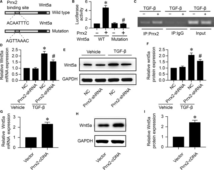
Figure 6.

Prrx2 functions as a transcriptional factor of Wnt5a which is up‐regulated by TGF‐β. A, The prediction of binding site for Prrx2 in Wnt5a promoter. B, HEK293A cells were cotransfected with pGL3‐promotor vector constructs expressing WT‐Wnt5a‐promoter containing ACAATTTC or MT‐Wnt5a‐promoter including AGTTAAAC plus Prrx2 cDNA plasmid. Cells were subjected to detect the relative luciferase activity. N is 5 per group. *P < .05 vs WT‐Wnt5a‐promoter. # P < .05 vs WT‐Wnt5a‐promoter plus Prrx2. C, Cultured cardiac fibroblasts were treated with TGF‐β (10 ng/mL) for 24 h. Cells were subjected to detect the binding of Prrx2 to Wnt5a gene promoter by using ChIP method. For ChIP experiment, complex of chromatin/protein was pulled down by Prrx2 primary antibody. The promoter of Wnt5a was amplified by PCR. Positive control is the 10% of the total chromatin in the absence of immunoprecipitation. Negative control is the chromatin immunoprecipitated with IgG and amplified with Wnt5a promoter primers. The PCR production is 200 bp. D‐F, Cultured cardiac fibroblasts were infected with adenovirus expressing negative control (NC) shRNA or Prrx2 shRNA for 48 h and then treated with TGF‐β (10 ng/mL) for 24 h. Total cell lysates were subjected to detect gene expression of Wnt5a by real‐time PCR in D and protein level of Wnt5a by Western blotting in E. Quantitative analysis of Wnt5a protein level was performed in F. N is 5 in each group. *P < .05 vs NC plus Vehicle. # P < .05 vs NC plus TGF‐β. G‐I, Cardiac fibroblasts were infected with adenovirus vector or expressing Prrx2 cDNA for 48 h. Cells were subjected to detect Wnt5a gene expression in G and protein level in H, and quantitative analysis of Wnt5a protein level was shown in I. N is 5 in each group. *P < .05 vs Vector
