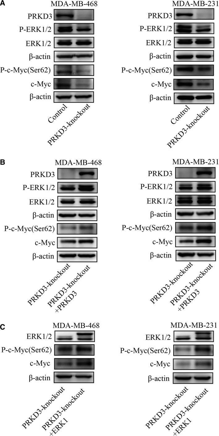
Figure 2.

Western blot analysis showed changes in the protein levels among PRKD3, (p‐)ERK1/2 and (p‐)c‐MYC. A, The protein levels of p‐ERK1 (Thr202/Tyr204), p‐c‐MYC (Ser62) and c‐MYC in the PRKD3‐knockout breast cancer cell lines were lower than the ones of these proteins in the parental cell lines (MDA‐MB‐468 and MDA‐MB‐231). B, Ectopic (over)expression of PRKD3 or (C) ERK1 in the PRKD3‐knockout cells led to the increased protein levels of (p‐)c ‐MYC(Ser62)
